Aortic image analyzing method and system

An analysis system and analysis method technology, applied in the field of aortic image analysis methods and systems, can solve the problems of high mortality, easy omission of details, fatigue, etc., reduce measurement errors, avoid the influence of human subjective factors, and realize automation and The effect of quickening

Inactive Publication Date: 2018-11-06
于存涛 +1
View PDF6 Cites 9 Cited by
  • Summary
  • Abstract
  • Description
  • Claims
  • Application Information

AI Technical Summary

Problems solved by technology

[0002] Aortic dissection is a serious clinical emergency with a high mortality rate, and the incidence rate has been on the rise in recent years. The diagnosis mainly relies on CTA. More importantly, the treatment of aortic dissection is very dependent on the measurement results of CTA, such as the location of the primary tear , the position of the secondary breach, the length of the dissection, the diameter, the accumulation of branch vessels, etc., but the current measurement method is manual measurement. Even for experienced measurers, it takes an average of 30 minutes, which is easy to cause fatigue. Details (such as the location of small secondary breaches) are easy to miss, and as the workload increases, the above problems become more obvious

Method used


Image

  • Aortic image analyzing method and system

Examples


Embodiment Construction

[0030] The specific implementation manners of the present invention will be further described in detail below in conjunction with the accompanying drawings and embodiments. The following examples are used to illustrate the present invention, but are not intended to limit the scope of the present invention.

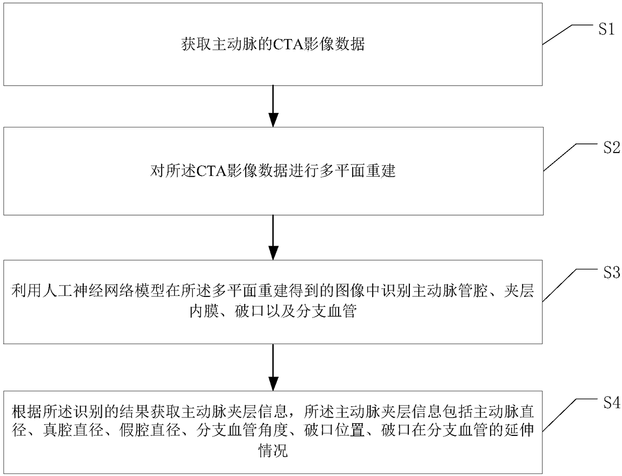
[0031] see figure 1 , figure 1 It is a flowchart of an aortic image analysis method provided in an embodiment of the present invention, including:

[0032] Step S1: acquiring CTA image data of the aorta;

[0033] Specifically, the DICOM data of the aorta, that is, the CTA image data of the aorta, can be obtained by performing a CTA scan on the human body;

[0034] Step S2: performing multiplanar reconstruction (MPR) on the CTA image data;

[0035] Wherein, this step can be realized by using an existing method, which will not be described in detail in the present invention;

[0036] Step S3: using the artificial neural network model to identify the aortic lumen, dissec...


PUM


Abstract

The present invention discloses an aortic image analyzing method and system. The aortic image analyzing method comprises the following steps: step S1: acquiring CTA image data of the aorta; step S2: performing multiplanar reconstruction on the CTA image data; step S3: utilizing an artificial neural network model to identify the aortic lumen, the intima of the interlayer, the crevasse, and the branch vessel in the image obtained through multiplanar reconstruction; step S4: acquiring aortic dissection information according to the result of identification, wherein the aortic dissection information comprises the aortic diameter, the true lumen diameter, the false lumen diameter, the branch vessel angle, the location of the crevasse, and the extension status of the crevasse in the branch vessel. The aortic image analyzing method provided by the invention facilitates achievement of automation and rapidity of diagnosis and measurement of the aortic dissection, which not only can reduce the manual workload, but also can reduce the measurement error and avoid the influence of subjective factors.

Description

technical field [0001] The invention relates to the technical field of medical image processing, in particular to an aortic image analysis method and system. Background technique [0002] Aortic dissection is a serious clinical emergency with a high mortality rate, and the incidence rate has been on the rise in recent years. The diagnosis mainly relies on CTA. More importantly, the treatment of aortic dissection is very dependent on the measurement results of CTA, such as the location of the primary tear , the position of the secondary breach, the length of the dissection, the diameter, the accumulation of branch vessels, etc., but the current measurement method is manual measurement. Even for experienced measurers, it takes an average of 30 minutes, which is easy to cause fatigue. Details (such as the location of small secondary breaches) are easy to miss, and as the workload increases, the above problems become more obvious. Contents of the invention [0003] The purpos...

Claims


Application Information

Patent Timeline
Patent Type & Authority Applications(China)
IPC IPC(8): G06K9/00
CPCG06V40/10G06V40/14
Inventor 于存涛吴进林丘俊涛邱家伟姜文祥刘燊
Owner 于存涛
Why Eureka
Try Eureka