
Three-dimensional MRI semi-automatic lesion image segmenting method and system

An image segmentation, semi-automatic technology, applied in the field of medical image processing, can solve the problems of failure to use the similarity of lesion shape and outline, the segmentation effect cannot reach clinical application, increase the number of misclassified voxels, etc., to improve the segmentation effect, segmentation The effect is good and the calculation amount is reduced

Active Publication Date: 2018-11-20
卢龙 +1
View PDF12 Cites 6 Cited by
  • Summary
  • Abstract
  • Description
  • Claims
  • Application Information

AI Technical Summary

Problems solved by technology

Therefore, classifying voxels on slices that do not contain lesions not only increases the number of misclassified voxels, but also reduces the efficiency of the method
[0008] (3) Failure to take advantage of the similarity of the shape and outline of lesions on adjacent MRI slices
[0009] (4) Failure to take advantage of the characteristics of the cluster distribution of lesion voxels, that is, the voxels around the lesion voxels are likely to be lesion voxels
[0010] (5) The voxels of the training classifier and the voxels to be classified by the classifier come from different subjects. Due to the differences between the subjects, the lesion voxels of the subjects to be segmented have some lesion voxels of the training subjects The characteristics that do not have, resulting in unsatisfactory classification results
[0013] (1) In MRI, the inside of the lesion has strong inhomogeneity, and the outside of the lesion is connected with high-intensity signals such as white matter, so that the segmentation effect of the current automatic segmentation method based on machine learning cannot reach the level of clinical application standard

Method used


Image

  • Three-dimensional MRI semi-automatic lesion image segmenting method and system
  • Three-dimensional MRI semi-automatic lesion image segmenting method and system
  • Three-dimensional MRI semi-automatic lesion image segmenting method and system

Examples


Embodiment Construction

[0070] In order to make the object, technical solution and advantages of the present invention more clear, the present invention will be further described in detail below in conjunction with the examples. It should be understood that the specific embodiments described here are only used to explain the present invention, not to limit the present invention.

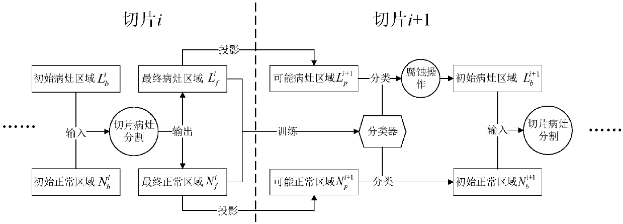
[0071] figure 1 , the three-dimensional MRI semi-automatic lesion image segmentation method provided by the embodiment of the present invention,

[0072] Step 1: Manual operation. The operator looks at the 3D MRI image to determine three things:

[0073] Information 1: The scope of the lesion slice [i min ,i max ], i.e. determine which slices are present with lesions in the whole brain;

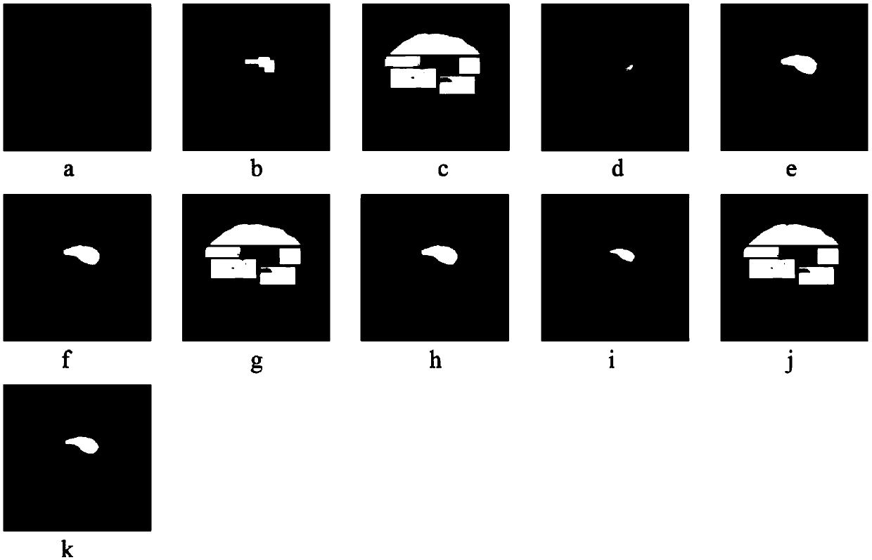
[0074] Information 2: the position i of the initial slice, select a slice from all lesion slices as the initial slice (see image 3 a), the selection criteria are as follows:

[0075] 1) The lesion area in the initial slice is larg...


PUM


Abstract

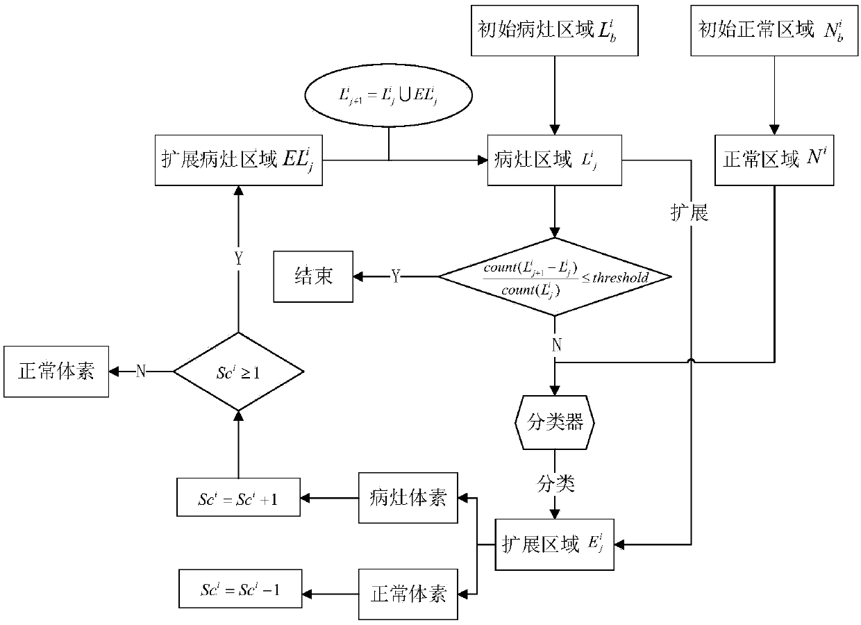
The invention belongs to the technical field of medical image processing, and discloses a three-dimensional MRI semi-automatic lesion image segmenting method and system. The method includes: determining a range of a lesion slice and a position of an initial slice by observing an MRI three-dimensional image, and dividing an initial lesion region and an initial normal region on the initial slice; segmenting the initial slice lesion, using a classifier to classify voxels in an extended region, and obtaining a final lesion region and a final normal region of the initial slice after multiple iterations; projecting the final lesion region and the final normal region on the initial slice onto an adjacent slice to obtain an initial lesion region and an initial normal region of the adjacent slice;and segmenting other section lesions, repeating slice lesion segmentation and region projection, segmenting the lesions within the lesion slice range, and performing combination to obtain the entire lesion region. After the above operation, the method can obtain a three-dimensional MRI lesion image.

Description

technical field [0001] The invention belongs to the technical field of medical image processing, and in particular relates to a three-dimensional MRI semi-automatic lesion image segmentation method and system. Background technique [0002] At present, the existing technologies commonly used in the industry are as follows: [0003] Magnetic resonance imaging (MRI) can clearly display soft tissue structures, and at the same time distinguish various tissues and lesions well, so it has been successfully applied to the diagnosis and treatment of various systems of the body. For the detection and treatment of various diseases, such as reperfusion therapy for cerebral apoplexy, early detection and radiation therapy of breast cancer, lung cancer and other cancers, it is necessary to quickly and accurately segment the lesion area, but manual segmentation of the lesion requires a lot of money It takes a lot of time and energy, and the accuracy of segmentation is affected by subjecti...

Claims


Application Information

Patent Timeline
IPC IPC(8): G06T7/00G06T7/11G06T7/12G06K9/62
CPCG06T7/0012G06T7/11G06T7/12G06T2207/10088G06T2207/20081G06T2207/20084G06F18/24
Inventor 胡联亭卢龙
Owner 卢龙
Learn More