
Sample preparation method for electron microscope scanning

A technology of sample preparation and electron microscope scanning, which is applied to the use of wave/particle radiation for material analysis, measuring devices, instruments, etc., can solve the problems of inability to provide mosquito and related insect electron microscope samples of Anopheles sinensis morphological classification, poor preparation performance, etc. Achieve the effect of avoiding deformation and comprehensive technology

Inactive Publication Date: 2019-01-18
CHONGQING NORMAL UNIVERSITY
View PDF3 Cites 1 Cited by
  • Summary
  • Abstract
  • Description
  • Claims
  • Application Information

AI Technical Summary

Problems solved by technology

[0008] The sample preparation performance of the prior art is poor; the morphological classification of mosquitoes generally not only relies on the morphological characteristics of adults, but also observes the morphological characteristics of each developmental stage for more accurate identification and classification, so as to accurately identify the species;
[0009] In the prior art, there is no electron microscope scanning and photographing and morphological description of the morphological characteristics of Anopheles sinensis at each developmental stage, which cannot provide a reference analysis method and data basis for the preparation of electron microscope samples of mosquitoes and related insects and the morphological classification of Anopheles sinensis

Method used


Image

  • Sample preparation method for electron microscope scanning
  • Sample preparation method for electron microscope scanning
  • Sample preparation method for electron microscope scanning

Examples


Embodiment Construction

[0044] In order to make the object, technical solution and advantages of the present invention more clear, the present invention will be further described in detail below in conjunction with the examples. It should be understood that the specific embodiments described here are only used to explain the present invention, not to limit the present invention.

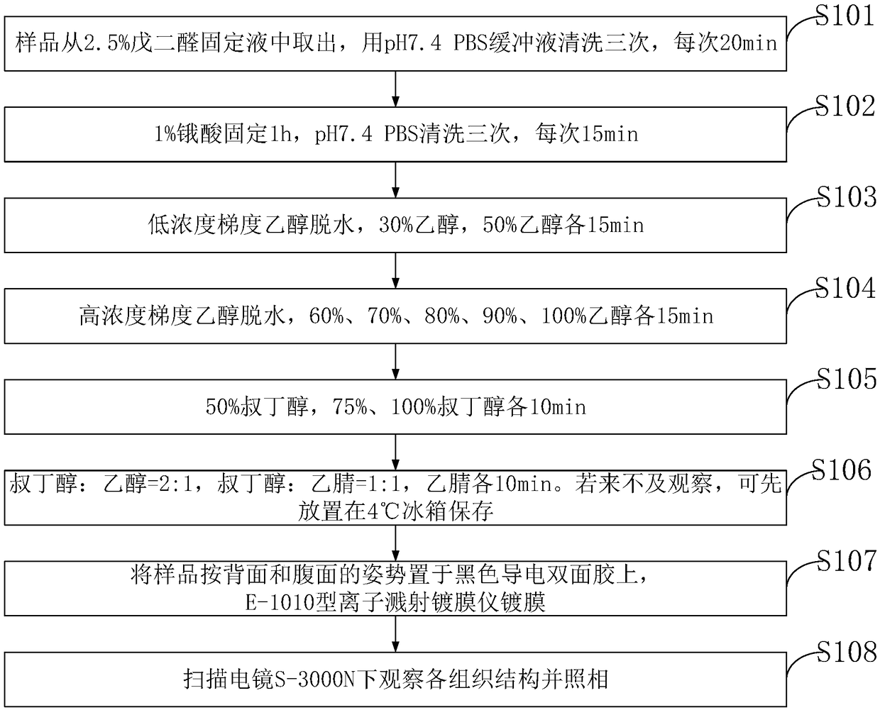
[0045] figure 1 , the sample preparation method for electron microscope scanning provided by the embodiment of the present invention includes:

[0046] S101: The sample is taken out from the 2.5% glutaraldehyde fixative, and washed three times with pH 7.4 PBS buffer, 20 min each time;

[0047] S102: fix with 1% osmic acid for 1 hour, wash with pH7.4 PBS three times, each time for 15 minutes;

[0048] S103: Dehydration with low-concentration gradient ethanol, 30% ethanol, 50% ethanol for 15 minutes each;

[0049] S104: Dehydration with high-concentration gradient ethanol, 60%, 70%, 80%, 90%, and 100% ethanol for 15 minute...


PUM


Abstract

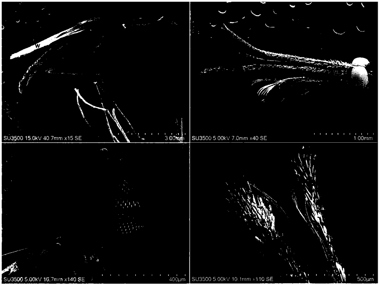
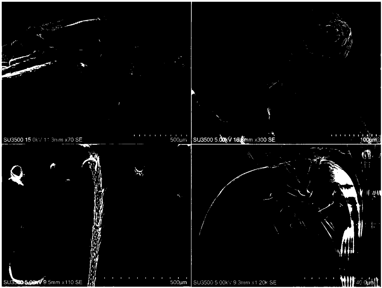
The invention belongs to the technical field of sample preparation, and discloses a sample preparation method for electron microscope scanning. The method comprises the following steps: taking out a sample from glutaraldehyde stationary liquid, and cleaning the sample for three times with a PBS buffer solution for 20 min each time; fixing the sample by uising 1% citric acid for 1 h, cleaning the sample for three times with the PBS with pH of 7 .4 for 15 min each time; performing dehydration by using low concentration gradient ethanol, and performing the dehydration by using 30% ethanol and 50%ethanol for 15 min respectively; and performing dehydration by using high concentration gradient ethanol. According to the sample preparation method disclosed by the invention, by exploring and improving the sample preparation technology of the electron scanning microscope (SEM), electron microscope samples of various developmental stages of mosquitoes are not only successfully prepared, electronmicroscope photographing and re-description are comprehensively performed on ultrafine structures of the anopheles sinensis at various developmental stages, thereby not only providing a more comprehensive technique for the preparation of the insect electron microscope samples, but also providing more detailed morphological characteristic data for the taxonomic analysis of the anopheles sinensis.

Description

technical field [0001] The invention belongs to the technical field of electron microscope materials, in particular to a sample preparation method for electron microscope scanning. Background technique [0002] At present, the existing technologies commonly used in the industry are as follows: [0003] The morphological classification of mosquitoes generally not only relies on the morphological characteristics of adults, but also observes the morphological characteristics of each developmental stage for more accurate identification and classification, so as to accurately identify the species. [0004] Scanning electron microscope (SEM) is an important tool in ultra-morphological analysis. SEM has the advantages of high resolution, large field of view, long depth of field and strong stereoscopic effect, and can conduct in-depth analysis of structures that are difficult to distinguish under an optical microscope. However, due to the small size of mosquitoes, it is difficult ...

Claims


Application Information

Patent Timeline
IPC IPC(8): G01N23/2202
CPCG01N23/2202
Inventor 闫振天陈斌
Owner CHONGQING NORMAL UNIVERSITY
Learn More