
A computer image processing method and apparatus for medical CT

An image processing and computer technology, applied in computer parts, computer-aided medical procedures, computing, etc., can solve problems affecting early diagnosis of diseases, slow image reconstruction speed, system errors, etc., to improve utilization rate, save time, The effect of improving efficiency

Active Publication Date: 2019-01-22
TIANJIN UNIV
View PDF6 Cites 8 Cited by
  • Summary
  • Abstract
  • Description
  • Claims
  • Application Information

AI Technical Summary

Problems solved by technology

[0003] In fact, CT images are reconstructed from the projection data of X-ray scanning of the human body. The reconstruction process will produce certain data loss and system errors, and the high-precision image reconstruction speed is slow, which affects the early diagnosis of diseases.

Method used


Image

  • A computer image processing method and apparatus for medical CT
  • A computer image processing method and apparatus for medical CT

Examples


Embodiment Construction

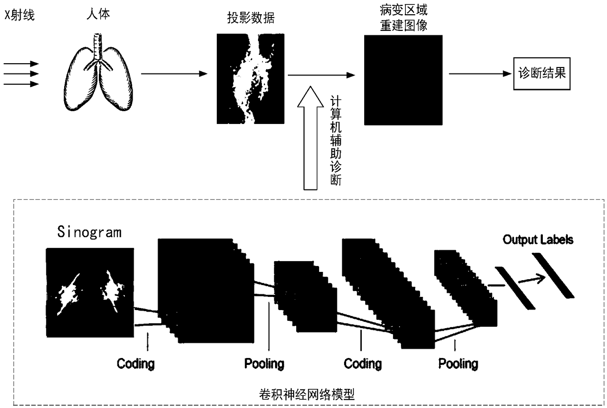
[0018] Aiming at the problem of missed diagnosis and misdiagnosis during computer-aided diagnosis, the present invention proposes a computer-aided diagnosis method for medical CT, such as figure 1 shown. The convolutional neural network is used to directly extract the features of the X-ray projection data through the human body and output the diagnostic results and the reconstructed image of the suspected lesion area, which improves the utilization rate of the projection data information and highlights the location of the lesion, which is beneficial to doctors. The second diagnosis has promoted the further development of precision medicine.

[0019] The present invention proposes a computer-aided diagnosis method for medical CT. First, a set of projection data is obtained through computerized tomography, and then it is used as input data through a convolutional neural network model to obtain a diagnosis result and a reconstructed image of a suspected lesion area. , the frame ...


PUM


Abstract

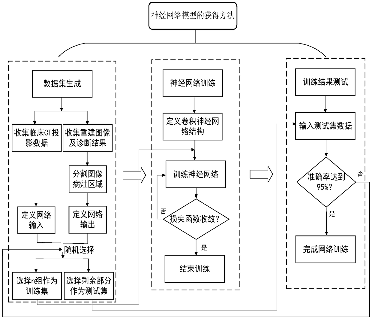
The invention relates to the field of computer-aided diagnosis, in order to improve the utilization rate of projection data information, and at the same time highlight the lesion position, which is conducive for doctors to carry out secondary diagnosis, and promote the further development of precision medicine. For this reason, the technical proposal adopted by the invention is a computer image processing method oriented to a medical CT, which comprises the following steps: Step 1: constructing a data set; Step 2: partitioning the dataset; Step 3: training convolution neural network; Step 4: testing the network training effect. The invention is mainly applied to the design and manufacture of computer-aided diagnosis medical equipment.

Description

technical field [0001] The invention relates to the field of computer-aided diagnosis, which utilizes deep learning to directly extract features from projection data and perform diagnosis, only performs image reconstruction on suspected lesion areas, and improves the accuracy and speed of medical diagnosis. Specifically, it relates to a computer image processing method and device for medical CT. Background technique [0002] Computed tomography (CT) is one of the commonly used disease detection methods in the medical field today. However, when a large number of images provide more detailed and accurate diagnostic information, it also increases the workload of the doctor who reads the film. It is easy to cause missed diagnosis and Misdiagnosis, and the qualitative analysis of lesions by doctors with experience in film reading is highly subjective, so computer-aided diagnosis can not only provide radiologists with accurate quantitative analysis to make up for the defects of hu...

Claims


Application Information

Patent Timeline
IPC IPC(8): G06K9/32G16H50/20A61B6/00
CPCG16H50/20A61B6/5217G06V10/25G06V2201/03
Inventor 史再峰李金卓曹清洁高静罗韬
Owner TIANJIN UNIV
Learn More