Automatic placement method used for medical image system and medical image system

A medical imaging and automatic technology, applied in the fields of radiological diagnostic equipment, medical science, application, etc., can solve the problem of low positioning efficiency

Pending Publication Date: 2019-01-29
SHANGHAI UNITED IMAGING HEALTHCARE
View PDF6 Cites 26 Cited by
  • Summary
  • Abstract
  • Description
  • Claims
  • Application Information

AI Technical Summary

Problems solved by technology

[0006] The invention provides an automatic positioning method for a medical imaging system and a medical imaging system, which are...

Method used


Image

  • Automatic placement method used for medical image system and medical image system
  • Automatic placement method used for medical image system and medical image system
  • Automatic placement method used for medical image system and medical image system

Examples


Embodiment 1

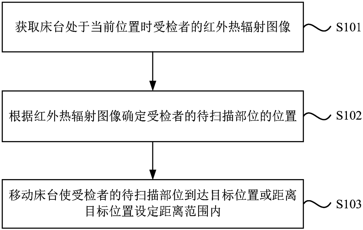
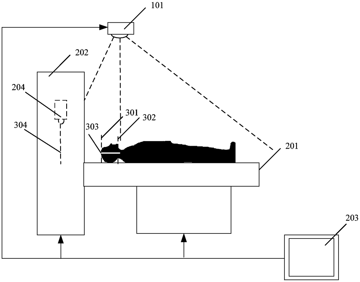
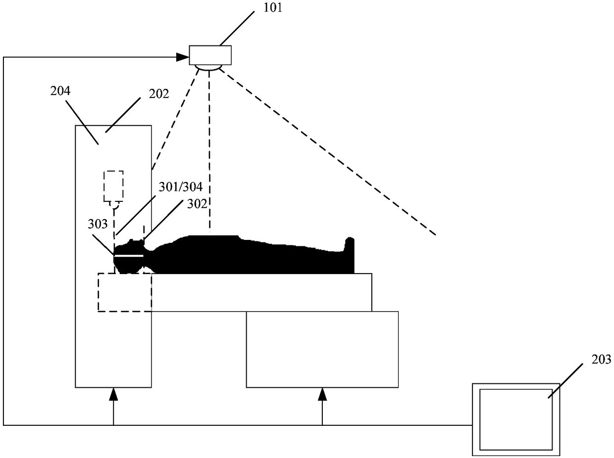
[0021] figure 1 It is a flowchart of an automatic positioning method for a medical imaging system provided by Embodiment 1 of the present invention. The technical solution of this embodiment is applicable to the automatic positioning of the medical imaging system before image scanning. Such as figure 2 and image 3 As shown, the medical imaging system includes a bed 201, a scanning device 202, a control device 203 connecting the bed 201 and the scanning device 202, and a photographing device 101 arranged above and / or on the side of the bed 201. The photographing device 101 It is fixed on the top of the image scanning room and includes an infrared detection device. The method specifically includes the following steps:

[0022] S101. Obtain an infrared thermal radiation image of the subject when the bed is at the current position.

[0023] In order to make the medical image have a higher clinical guiding significance, the technician needs to adjust the horizontal position a...

Embodiment 2

[0039] Such as figure 2 As shown, the medical imaging system includes a scanning device 202, and a bed 201 that moves axially along the scanning aperture of the scanning device 202, and also includes: a control device 203 and an imaging device 101 disposed above and / or on the side of the bed 201, The photographing device 101 includes an infrared detection device, which is used to obtain an infrared thermal radiation image of the subject; the control device 203 is connected to the photographing device in communication, and is used to control the movement of the bed platform 201 according to the infrared thermal radiation image of the subject, so that the subject The part to be scanned moves to the target position or within a set distance from the target position.

[0040] Wherein, the control device 203 includes an image processing module, and through the image processing module, according to the infrared thermal radiation image of the subject and the preset scanning protocol ...

Embodiment 3

[0049] Embodiment 3 of the present invention also provides a storage medium containing computer-executable instructions, the computer-executable instructions are used to execute an automatic positioning method for a medical imaging system when executed by a computer processor, the method include:

[0050] Obtain the infrared thermal radiation image of the subject when the bed is at the current position;

[0051] determining the position of the part to be scanned of the subject according to the infrared thermal radiation image;

[0052] The bed is moved so that the part to be scanned of the subject arrives at a target position or within a set distance from the target position.


PUM


Abstract

The invention discloses an automatic placement method used for a medical image system and the medical image system. The medical image system includes a scanning device, a bed platform axially moving along scanning apertures of the scanning device and a photographing device arranged at the upper portion and/or lateral portion of the bed platform. The photographing device includes an infrared detector. The method includes the steps of obtaining an infrared thermal radiation image of a subject when the bed platform stays at the current position; according to the infrared thermal radiation image,determining a to-be-scanned part of the subject; moving the bed platform to make the to-be-scanned part of the subject reach a target position or positioned within a set distance range from the targetposition. The method solves the problem that existing placement methods in the prior art are low in efficiency, and has the technical advantage that the placement efficiency is improved.

Description

technical field [0001] Embodiments of the present invention relate to medical equipment technology, and in particular to an automatic positioning method for a medical imaging system and the medical imaging system. Background technique [0002] Modern medical imaging technology mainly includes X-ray imaging (X-ray Radiography, referred to as XR), computed tomography (Computed Tomography, referred to as CT), magnetic resonance tomography (Magnetic Resonance, referred to as MR), positron emission computed tomography (Positron Emission Tomography-Computed Tomography (PET-CT for short), and the latest Positron Emission Magnetic Resonance Tomography (PET-MR for short); and Accelerator Radiation Therapy (RT for short) is one of the modern tumor treatment technologies. [0003] In order to accurately determine the scanning range of the human body, before the CT, MR, PET-CT or PET-MR system scans the specific organs of the human body, it is first necessary to obtain a topogramm throu...

Claims


Application Information

Patent Timeline
IPC IPC(8): A61B5/055A61B6/04A61N5/10
CPCA61N5/1069A61B6/0407A61N2005/1097A61B6/0487A61B5/055
Inventor 胡仁芳
Owner SHANGHAI UNITED IMAGING HEALTHCARE
Why Eureka
Try Eureka