Pepsinogen II detection kit

A pepsinogen and detection kit technology, applied in biological testing, measurement devices, material inspection products, etc., can solve problems such as low anti-interference performance, long measurement time, interference with test results, etc., to improve anti-interference ability, Eliminate the effect of heterophilic antibodies

Inactive Publication Date: 2019-02-05
AUTOBIO DIAGNOSTICS CO LTD
View PDF2 Cites 7 Cited by
  • Summary
  • Abstract
  • Description
  • Claims
  • Application Information

AI Technical Summary

Problems solved by technology

The enzyme-linked immunosorbent assay method is complicated to operate, takes a long time to measure, has many influencing factors, and is not suitable for the detection of emergency samples; when the amount of antigen or antibody is too large, soluble complexes may appear in the immunoturbidimetric method, causing errors. susceptible to lipemia
Although the chemiluminescent immunoassay method has many advantages such a

Method used


Image

  • Pepsinogen II detection kit
  • Pepsinogen II detection kit
  • Pepsinogen II detection kit

Examples


Embodiment 1

[0017] Example 1 Preparation of Pepsinogen II Detection Kit

[0018] 1. Preparation of magnetic particle suspension

[0019] After fully mixing and washing the magnetic particle stock solution with a particle size of 1.08 μm, add 10-50 mg / ml of glutaraldehyde activator, mix and shake, and after washing, add 0.1-0.5 μg / person of pepsinogen Ⅰ Antibody, mix and shake, and finally block with PBS and 1%~5% bovine serum albumin blocking solution, and store at 2-8°C.

[0020] 2. Preparation of Enzyme Diluent

[0021] Add 1%~5% bovine serum albumin to the prepared MES buffer, mix it to become the enzyme diluent, add the HRP-labeled antibody according to the ratio of 1:500~1:5000 and mix well, 2~8℃ save.

[0022] 3. Preparation of calibrator

[0023] Add 1%~5% bovine serum albumin to the prepared MOPS buffer, mix well to obtain the calibrator diluent, dilute the PGⅡ antigen into 6 gradients with the calibrator diluent, 0ng / ml, 10ng / ml, 25ng / ml, 50 ng / ml, 100 ng / ml, 200 ng / ml, aliq...

Embodiment 2

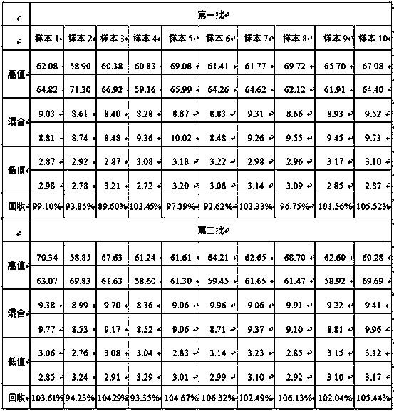
[0027] Example 2 Evaluation of sample storage conditions

[0028] Take 10 cases of fresh samples of pepsinogen II gradient, and test them at 0h, 8h, 2d, 3d, and 5d respectively, and compare the test results at each time with the 0h results to assess the stability of the samples. Samples were stored at 2-8°C during the experiment. The results are shown in Table 2 below:

[0029] Table 2 Research on the storage time of the kit samples of the present invention

[0030]

[0031] The results in Table 2 show that the storage time of the samples of the kit of the present invention is up to 5 days at 2-8°C.

Embodiment 3

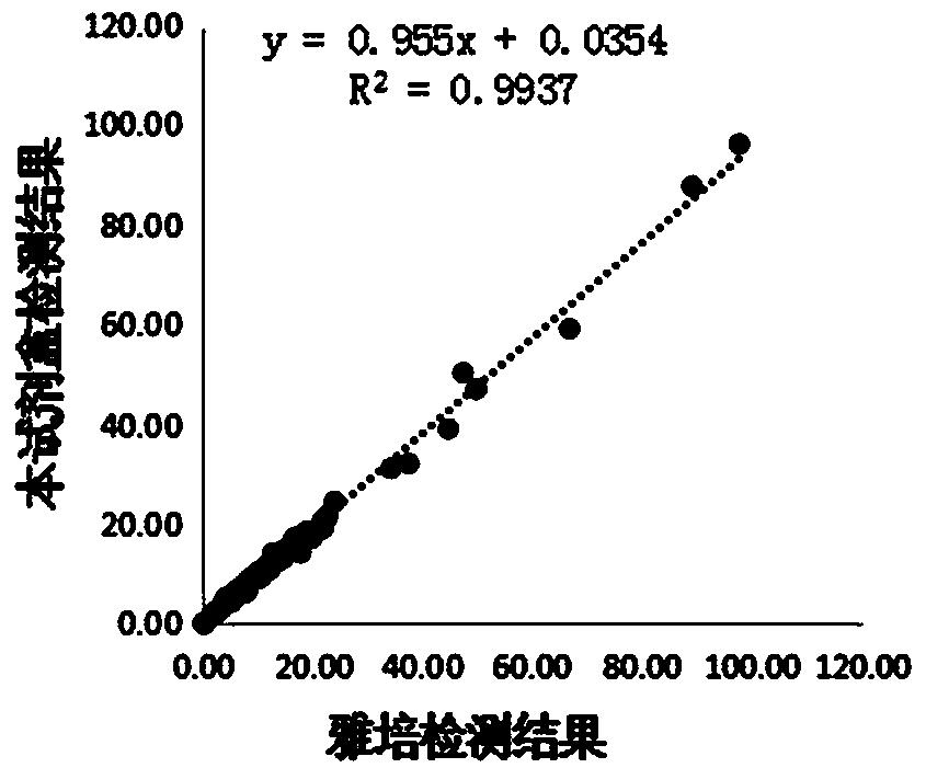
[0032] Embodiment 3 Performance evaluation of the kit of the present invention

[0033] 1. Sensitivity detection

[0034] LOB, prepare 5 clinical samples with a value close to 0, repeat 3 times for each sample, and do a total of 4 days to obtain 60 data; LOD, prepare 5 serial clinical samples with a concentration range of 1-4 times LOB, each sample Repeat 3 times, do a total of 4 days, and get 60 data; FS: Using the data in the LOD experiment, 5 concentration samples are measured 3 times a day, a total of 4 days, each sample gets 12 results, and the calculation of each sample Mean, SD and CV%, the concentration closest to 20% is the functional sensitivity; the test results of the two batches are shown in Table 3.

[0035] Table 3 Sensitivity detection of the kit of the present invention

[0036]

[0037] It can be seen from the results in Table 3 that the concentration that can be accurately detected in the first batch is 0.85 ng / mL, and the concentration that can be accu...


PUM

PropertyMeasurementUnit
Particle sizeaaaaaaaaaa

Abstract

The invention discloses a pepsinogen II detection kit. The kit comprises a magnetic particle coated with a pepsinogen II monoclonal antibody, an enzyme diluent containing a horseradish peroxidase (HRP) labeled pepsinogen II monoclonal antibody, and a calibrator and a sample diluent containing a pepsinogen II antigen. A blocker component is contained in the sample diluent. The blocker is one or more human anti-mouse antibodies which neutralizes interfere of the HAMA. The method has the advantages that the high-sensitivity chemiluminescence technology and the magnetic particle preparation technology are combined, and an AutoLumo full-automatic detection analyzer is used in cooperation, so that a full-automatic detection is realized; meanwhile, the blocker component is added into the sample diluent, so that the heterophile antibody in the sample can be eliminated, and the anti-interference capability of the kit is greatly improved.

Description

technical field [0001] The invention relates to biological detection technology, in particular to a pepsinogen II detection kit. Background technique [0002] Pepsinogen (PG) is the inactive precursor of pepsin (proteolytic enzyme) in gastric juice. It is a protein polypeptide chain composed of 375 amino acids, with an average relative molecular mass of 42000 Da. There are 7 groups of pepsin in human gastric mucosa isoenzymes. According to biochemical properties, immunogenicity, cell source and tissue distribution, it can be divided into two subgroups, pepsinogen Ⅰ (PGⅠ) and pepsinogen Ⅱ (PGⅡ). Components 1-5 have similar immunogenicity and are called PGⅠ. , mainly secreted by the mucous neck cells of the principal cells of the fundic glands; components 6-7 are called PGⅡ, in addition to being secreted by the chief cells of the oxyntic glands of the gastric body and fundic mucosa, the mucous neck cells of the oxyntic glands PGII can also be produced by the mucus cells of t...

Claims


Application Information

Patent Timeline
IPC IPC(8): G01N33/577G01N33/573G01N33/535G01N33/68G01N21/76
Inventor 马雷史小芹渠文涛刘雅奇周金龙田会会李晓霞乔晓芳付光宇吴学炜
Owner AUTOBIO DIAGNOSTICS CO LTD
Why Eureka
Try Eureka