Chimeric antigen receptor DAP12-T2A-CD8alpha-MSLN scFv-NKp44 and purpose thereof

A technology of DAP12-T2A-CD8, -mslnscfv-nkp44, applied in the field of immune cells, can solve the problem of cytokine release syndrome without observed acute adverse events

Inactive Publication Date: 2019-03-15
NANJING CART MEDICAL TECH LTD
View PDF2 Cites 2 Cited by
  • Summary
  • Abstract
  • Description
  • Claims
  • Application Information

AI Technical Summary

Problems solved by technology

No infusion-related acute adverse events (AEs) and cytokine release syndrome were observed in six patients with recurrent serous ovaria...

Method used


Image

  • Chimeric antigen receptor DAP12-T2A-CD8alpha-MSLN scFv-NKp44 and purpose thereof
  • Chimeric antigen receptor DAP12-T2A-CD8alpha-MSLN scFv-NKp44 and purpose thereof
  • Chimeric antigen receptor DAP12-T2A-CD8alpha-MSLN scFv-NKp44 and purpose thereof

Examples


Embodiment 1

[0050] Embodiment 1, chimeric antigen receptor preparation

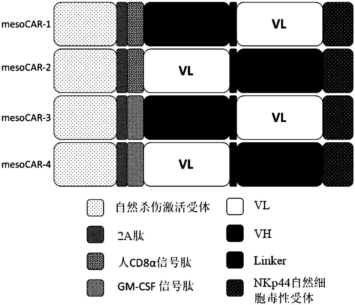
[0051] The present invention provides an optimized chimeric antigen receptor targeting MSLN antigen. The chimeric antigen receptor of the present invention is composed of the second intracellular conduction domain-T2A-extracellular signal peptide-targeting MSLN antigen binding domain-the first intracellular conduction domain in series. Therefore, viral vectors containing different combinations of stimulation signals need to be constructed separately. In this example, NKp44 is used as the unified structure of the first intracellular conduction domain, and the following four chimeric antigen receptors need to be constructed respectively ( figure 1 ):

[0052] DAP12-T2A-CD8α signal peptide-VH-Linker-VL-NKp44 (mesoCAR-1)

[0053] DAP12-T2A-CD8α signal peptide-VL-Linker-VH-NKp44 (mesoCAR-2)

[0054] DAP12-T2A-GM-CSF signal peptide-VH-Linker-VL-NKp44 (mesoCAR-3)

[0055] DAP12-T2A-GM-CSF signal peptide-VL-Linker-VH-NK...

Embodiment 2

[0101] Embodiment 2, virus infection T cell

[0102] 1. Isolation and activation of T cells and virus infection

[0103] (1) Isolation of human peripheral blood mononuclear cells

[0104] Use a blood collection tube containing anticoagulant to collect about 10ml of peripheral blood, settle naturally at room temperature (18-25°C) for about 30min, collect the upper layer of plasma, and centrifuge the collected upper layer of plasma at 5000r / min for 10min at a volume ratio of 1:1 Add to the lymphocyte separation medium (purchased from Tianjin Haoyang Biological Products Technology Co., Ltd.), gradient centrifugation, 3000r / min, centrifugation for 30min, after centrifugation, the centrifuge tube is layered from top to bottom: the first layer is the plasma layer ; The second layer is the buffy coat layer of lymphocytes; the third layer is the transparent separation liquid layer; the fourth layer is the red blood cell layer. Aspirate the buffy coat of lymphocytes, wash twice with ...

Embodiment 3

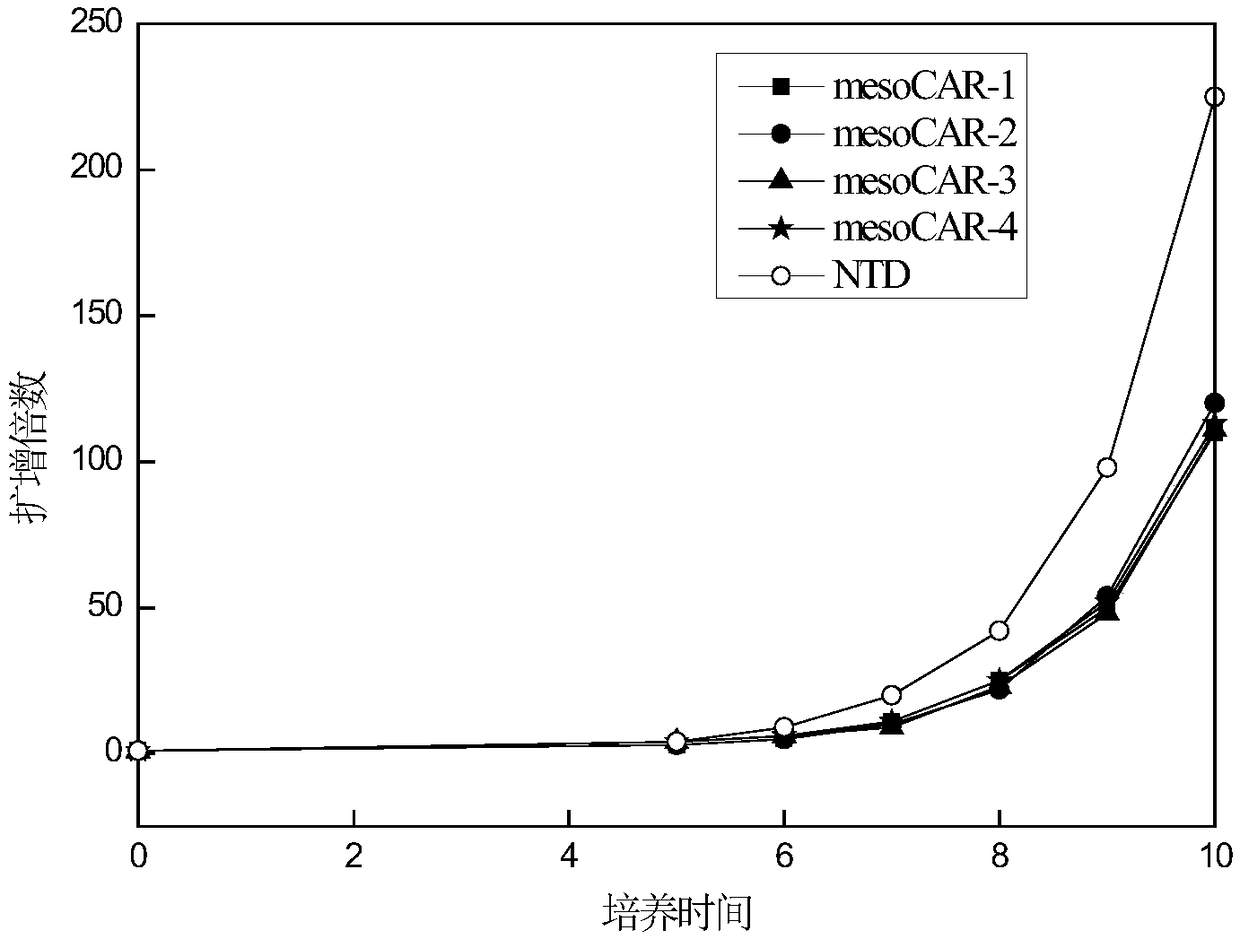
[0109] Example 3. Effect of Virus Infection of CAR-T Cells on Cell Proliferation

[0110] After each group of viruses infected T cells, the T cells were counted every 1-2 days with a complete medium containing 5% autologous plasma + 300 IU / ml recombinant human IL-2 + KBM581. Then observe the growth of T lymphocytes, the results are as follows: image 3 shown. The results showed that after the cells were infected with CAR-expressing viruses, they could still form typical proliferative clonal clusters. By counting the cells and drawing the cell proliferation curves, it could be seen that mesoCAR-1, mesoCAR-2, mesoCAR-3, and mesoCAR-4 proliferated similarly, compared with T cells uninfected with the virus ( image 3 Medium NTD) proliferative ability was slightly weaker.


PUM

PropertyMeasurementUnit
Titeraaaaaaaaaa

Abstract

The invention discloses a chimeric antigen receptor DAP12-T2A-CD8alpha-MSLN scFv-NKp44 and a purpose thereof. The chimeric antigen receptor DAP12-T2A-CD8alpha-MSLN scFv-NKp44 comprises an intracellular second transduction domain DAP12, T2A, a CD8alpha signal peptide, anti-human MSLN monoclonal antibody ss1 light chain and heavy chain variable regions MSLN scFv and an intracellular first transduction domain which are connected with each other in series. The chimeric antigen receptor DAP12-T2A-CD8alpha-MSLN scFv-NKp44 disclosed by the invention is used for modifying T lymphocytes; and the modified T cells (CAR-T cells) can be used for treating MSLN positive malignant solid tumors. In a malignant solid tumor killing test, the killing ability of the CAR-T cells to pancreatic tumor cells is obviously enhanced, and good safety and antineoplastic activity are shown in clinical application.

Description

technical field [0001] The invention relates to the technical field of tumor immunotherapy. The present invention uses genetic engineering technology to obtain a fusion gene encoding chimeric antigen receptor DAP12-T2A-CD8α-MSLN scFv-NKp44, inserts the gene fragment into a lentiviral expression vector, packages it into a lentivirus, and transduces it into human T lymphocytes. T cells are made to express the chimeric antigen receptor. The present invention also relates to nucleic acids encoding such transmembrane polypeptides, vectors, and immune cells expressing the CAR on their surface for immunotherapy. The chimeric antigen receptor-modified T cells involved in the present invention can target and specifically kill MSLN-positive tumor cells for the treatment of malignant solid tumors, and open up an effective adoptive immunotherapy strategy for the treatment of cancer path of. Background technique [0002] With the development of tumor immunology theory and clinical tec...

Claims


Application Information

Patent Timeline
IPC IPC(8): C07K19/00C12N15/62C12N15/867C12N5/10A61K35/17A61P35/00
CPCA61K35/17A61P35/00C07K14/70503C07K14/70517C07K14/7056C07K16/30C07K2317/622C07K2319/00C07K2319/02C07K2319/33C07K2319/74C12N5/0636C12N15/86C12N2510/00C12N2740/15043C12N2800/107
Inventor 王恩秀汪晨武国英
Owner NANJING CART MEDICAL TECH LTD
Why Eureka
Try Eureka