Endoscopic image processing method, apparatus, system, and storage medium

An image processing device and endoscope technology, applied in the field of image processing, can solve problems such as insufficient robustness, incomplete coverage, and high threshold, and achieve the effect of improving resource utilization, reducing dependence on professional level, and reducing quantity

Active Publication Date: 2019-03-26
腾讯医疗健康(深圳)有限公司
View PDF11 Cites 23 Cited by
  • Summary
  • Abstract
  • Description
  • Claims
  • Application Information

AI Technical Summary

Problems solved by technology

However, this existing technology requires researchers to deeply understand the medical image itself, and then formulate a usable feature extraction scheme based on the inherent characteristics of the image, which has a high threshold
In addition, the extracted features are more biased towards general features, not specific organ features for the specific body parts to be diagnosed, which makes the coverage incomplete and the robustness of the scheme is not good enough

Method used


Image

  • Endoscopic image processing method, apparatus, system, and storage medium
  • Endoscopic image processing method, apparatus, system, and storage medium
  • Endoscopic image processing method, apparatus, system, and storage medium

Examples


Embodiment Construction

[0030] In order to make the object, technical solution and advantages of the present invention clearer, the present invention will be further described in detail below with reference to the accompanying drawings and examples.

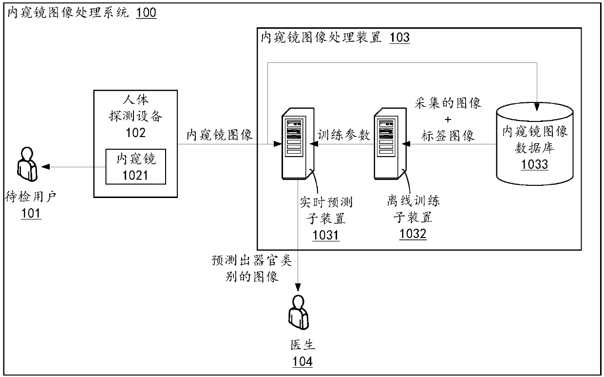
[0031] figure 1 It is a schematic structural diagram of an endoscope image processing system involved in an embodiment of the present invention. Such as figure 1 As shown, the endoscope image processing system 100 includes a user to be checked 101 , a human body detection device 102 including an endoscope 1021 , an endoscope image processing device 103 and a doctor 104 . Wherein, the endoscopic image processing device 103 includes a real-time prediction sub-device 1031 , an offline training sub-device 1032 and an endoscopic image database 1033 .

[0032] According to an embodiment of the present invention, the human body detection device 102 detects a certain body part of the user 101 to be checked through the endoscope 1021 . The human body detectio...


PUM


Abstract

Disclosed are an endoscopic image processing method, apparatus, system, and storage medium. The method includes: acquiring a first endoscopic image for a human body part; Creating a depth convolutionnetwork for predicting an endoscopic image, and determining training parameters of the depth convolution network based on the first endoscopic image and at least one second endoscopic image transformed from the first endoscopic image; And acquiring a current endoscopic image of a user to be examined, predicting the current endoscopic image based on a depth convolution network and training parameters, and determining an organ type corresponding to the current endoscopic image. The method of the invention makes the prediction process more intelligent and more robust, and improves the resource utilization rate of the processing device.

Description

technical field [0001] The present invention relates to the technical field of image processing, in particular to an endoscopic image processing method, device, system and storage medium. Background technique [0002] At present, gastric cancer and esophageal cancer rank among the top five types of malignant tumors in my country and the world. Gastric cancer and esophageal cancer are both malignant tumors of the upper gastrointestinal tract. In actual clinical practice, the doctor conducts electronic inspection through the endoscope, inserts the endoscope from the oral cavity into the upper digestive tract of the subject, and uses the strong light emitted by the light source to bend the light through the light guide fiber, so that the doctor can Observe the health status of various organs in the upper digestive tract. [0003] However, due to differences in the acquisition environment, detection equipment, and doctor’s shooting habits of medical images obtained through end...

Claims


Application Information

Patent Timeline
Patent Type & Authority Applications(China)
IPC IPC(8): G06T7/00G06N3/04
CPCG06T7/0012G06T2207/20081G06T2207/10068G06T2207/30004G06N3/045A61B1/0005G06N3/084A61B1/000096A61B1/000094
Inventor 付星辉孙钟前杨巍
Owner 腾讯医疗健康(深圳)有限公司
Why Eureka
Try Eureka