Multi-angle image system for thoracoscope and peritoneoscope

An imaging system and thoracoabdominal cavity technology, applied in the medical field, can solve problems such as inconvenient operation, increased patient pain, and misjudgment during operation, so as to increase the operating space and moving space, improve accuracy and success rate, and reduce The effect of wound infection

Pending Publication Date: 2019-04-05
福州骏格科技有限公司
View PDF11 Cites 1 Cited by
  • Summary
  • Abstract
  • Description
  • Claims
  • Application Information

AI Technical Summary

Problems solved by technology

This operation method must be held continuously or the endoscope lens must be clamped by external instruments, which is very inconvenient to operate
[0003] In addition, the laparoscope used in the existing thoracic and laparoscopic surgery cannot preview the images in the thoracoabdominal cavity, and can only manually adjust the shooting angle and position of the endoscopic lens to find the surgical area of ​​the lesion during the operation; moreover, the only The viewing angle is usually the forward viewing angle of the chief surgeon. The assistants are located on the opposite side of the chief surgeon to operate the instrument viewing angle, but they have to share the same endoscopi

Method used


Image

  • Multi-angle image system for thoracoscope and peritoneoscope
  • Multi-angle image system for thoracoscope and peritoneoscope
  • Multi-angle image system for thoracoscope and peritoneoscope

Examples


Embodiment 1

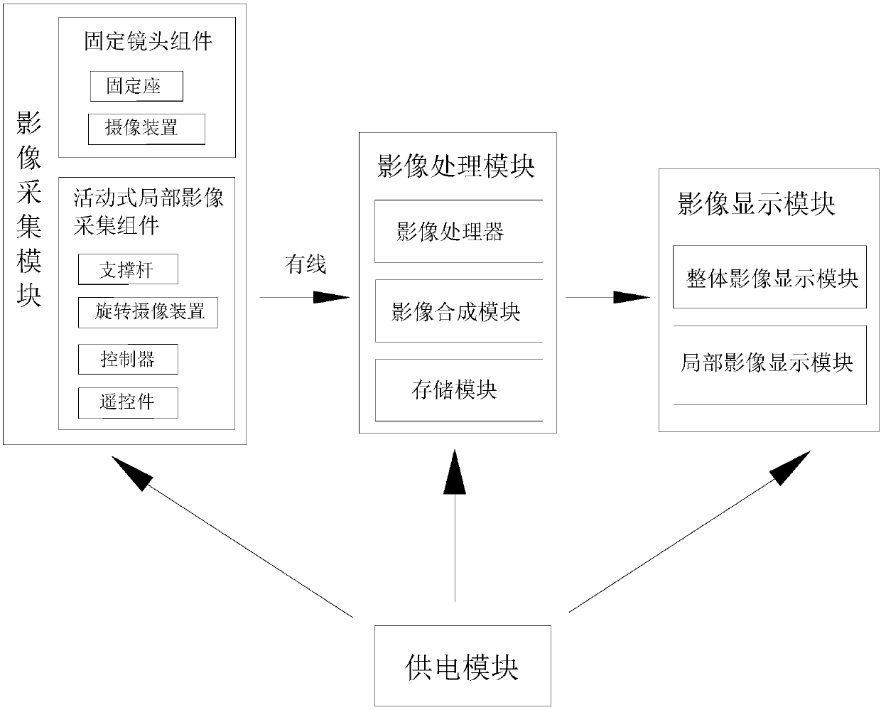
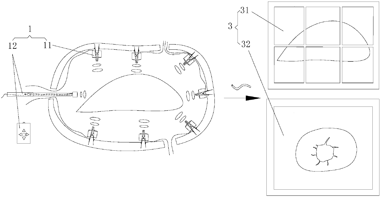
[0057] Such as Figure 1-Figure 10 Shown, a kind of multi-angle imaging system of thoraco-laparoscopy is characterized in that: comprising

[0058] The image acquisition module 1 is used to capture image data in the thoracic and abdominal cavity and send the image data signal to the outside;

[0059] The image processing module 2 is signal-connected with the image acquisition module 1, and is used to receive and process the image data signal transmitted by the image acquisition module 1;

[0060] The image display module 3 is connected to the output terminal of the image processing module 2, and is used to receive the processed image data and display it on the end surface of the screen;

[0061] The power supply module 4 is used to provide the electric energy required for the operation of each module;

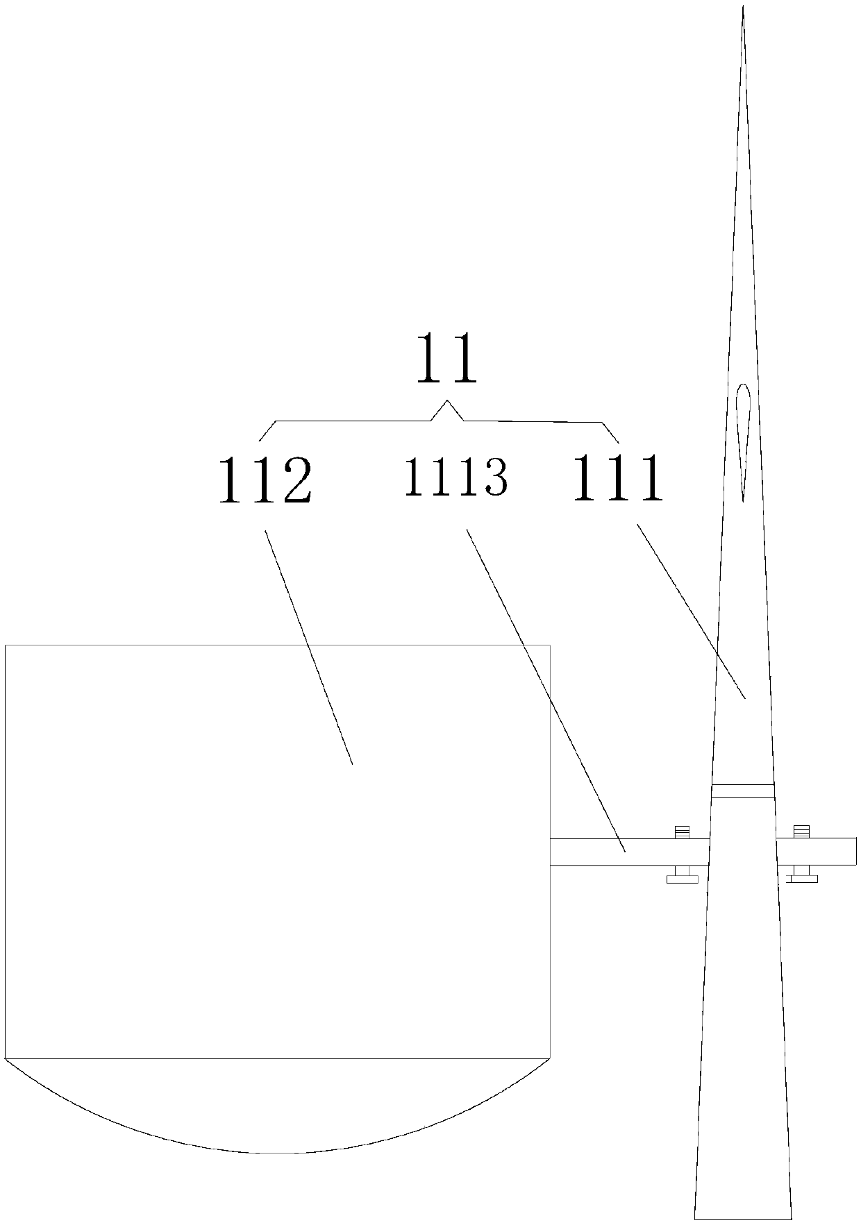
[0062] The image acquisition module 1 includes more than one set of fixed lens assemblies 11 that are fixedly connected to the inner side of the thoracic and abdominal cavity...

Embodiment 2

[0087] Such as Figure 11-Figure 14 As shown, the difference between the multi-angle image system of a kind of thoraco-laparoscopic in the present embodiment and embodiment 1 is:

[0088] The image acquisition module 1 and the image processing module 2 are connected by wireless signals.

[0089] When the image acquisition module 1 and the image processing module 2 are connected by wireless signal,

[0090] The fixed lens assembly 11 also includes a first wireless transmitter 113 for transmitting the image data captured by the camera 112; Two wireless transmitters 125;

[0091] The image processing module 2 is also provided with a wireless receiver 22 for receiving the image data signal transmitted by the wireless transmitter, and the output end of the wireless receiver 22 is connected with the input end of the image processor 21;

[0092] The power supply module 4 also includes a first battery assembly 41 disposed in the fixed lens assembly 11 to provide the working power r...


PUM


Abstract

The invention relates to the medical field and especially relates to a multi-angle image system for a thoracoscope and a peritoneoscope. The multi-angle image system is characterized by comprising animage collecting module, an image processing module, an image display module and a power supply module; the image collecting module comprises a fixed lens component and a mobile local image collectingcomponent; the fixed lens component comprises a fixed base and a camera device; the mobile local image collecting component comprises a support rod, a rotary camera device, a controller and a remotecontroller; an image processor is arranged in the image processing module; the image display module comprises a whole image display module and a local image display module. The multi-angle image system for the thoracoscope and the peritoneoscope is capable of comprehensively displaying surgical environment images in thoracic cavity and abdominal cavity; medical personnel can have a multi-angle macroscopical understanding for the environment in the thoracic cavity and the abdominal cavity and can observe dead angles of view of the present endoscope technology; doctors can conveniently operate on the affected parts and the accuracy and success rate of operation can be increased.

Description

technical field [0001] The invention relates to the medical field, in particular to a multi-angle imaging system for thoracic and laparoscopy. Background technique [0002] As a newly developed minimally invasive technique, laparoscopic surgery refers to making an incision on the affected area, holding the endoscopic lens and various special surgical instruments through the incision and inserting them into the patient's abdominal cavity. The images of various organs in the abdominal cavity captured by the endoscopic lens in the abdominal cavity are transmitted to the TV screen, and the surgeon completes the operation by observing the images and operating outside the body with various surgical instruments. This mode of operation must be held continuously or an external device is used to clamp the endoscopic lens, which is very inconvenient to operate. [0003] In addition, the laparoscope used in the existing thoracic and laparoscopic surgery cannot preview the images in the...

Claims


Application Information

Patent Timeline
IPC IPC(8): A61B1/313A61B1/00A61B1/05
CPCA61B1/00009A61B1/00016A61B1/00018A61B1/05A61B1/313
Inventor 王财生赵林栋
Owner 福州骏格科技有限公司
Why Eureka
Try Eureka