CT scanning method of low radiation dosage

A CT scanning, low-radiation technology, used in computed tomography scanners, echo tomography, etc., can solve the problems of CT image noise signal enhancement, improve lesions, image quality degradation, etc., to reduce X-ray radiation dose and improve quality. , the effect of fast reconstruction speed

Pending Publication Date: 2019-05-07
SHANGHAI JIAO TONG UNIV
View PDF3 Cites 4 Cited by
  • Summary
  • Abstract
  • Description
  • Claims
  • Application Information

AI Technical Summary

Problems solved by technology

One type of method is to reduce the intensity of X-rays to reduce the radiation dose, but the problem is that the noise signal of the CT image is relatively enhanced, and the image quality is degraded.
Considering that many lesions are small in size and rich in details, severe noise signals will greatly increase the difficulty of lesion detection and increase the risk of missed detection and false detection; another method is to reduce the radiation dose by reducing the X-ray projection beam, such as Reduce the number of projection angles
Many existing methods perform high-quality CT image reconstruction from a small amount of data with a limited number of angles

Method used


Image

  • CT scanning method of low radiation dosage
  • CT scanning method of low radiation dosage
  • CT scanning method of low radiation dosage

Examples


Embodiment 1

[0049] Target lesion: pulmonary nodule

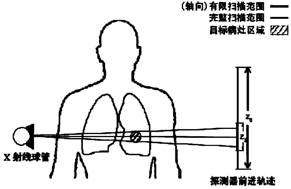
[0050] Scanning method: the scanning center is positioned near the nodules, the scanning range is preset, and the scanning covers the local lung area

[0051] Use the previous lung examination CT scan of the same patient to locate and scan the current lesion area. The specific steps are as follows:

[0052] Step 1. According to the position of the pulmonary nodule confirmed by the previous CT scan, the three-dimensional position of the target lesion is estimated and positioned. The doctor predicts the growth of the lesion to estimate the size of the nodule, and determines the reconstruction range required by the CT scan.

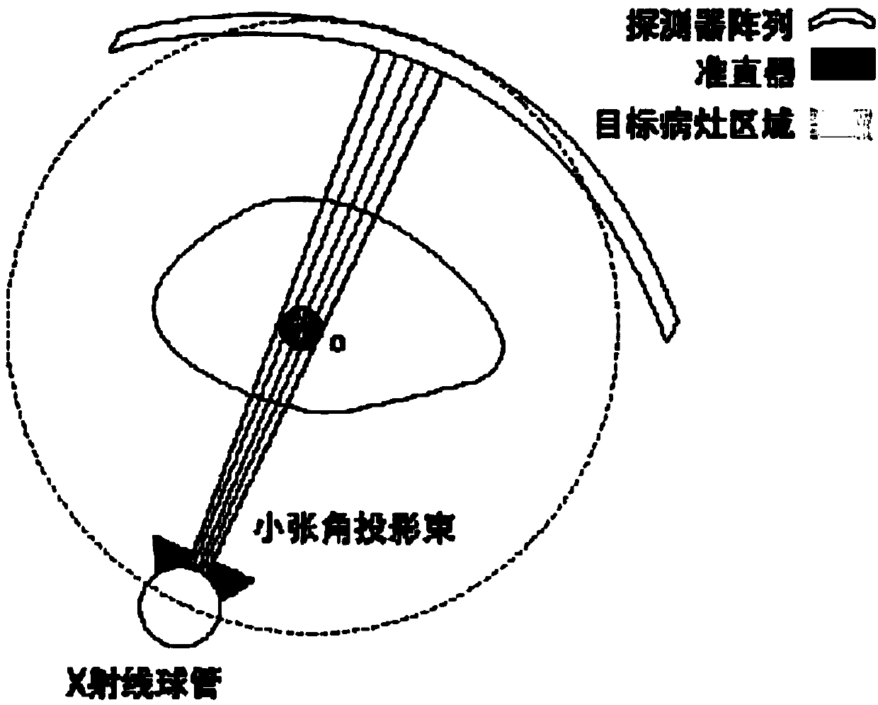
[0053] Step 2. Design a CT scanning method with a limited number of projections according to the staging situation and imaging requirements of the latest pulmonary nodules. In this embodiment, the scanning method can be designed from the following three aspects: limited cross-sectional design, first outline the circu...

Embodiment 2

[0058] Target lesion: intracranial calcification

[0059] Scanning method: The scanning center is positioned to the calcification point of the brain, and the scanning range and interval rotation angle are preset. The scan covers a localized brain area.

[0060] Also use the CT image of the same patient with previous calcified lesions to locate and scan the area near the target intracranial calcified point. The specific steps are as follows:

[0061] Step 1. The doctor locates the three-dimensional position of the target calcification, and estimates the reconstruction range of the CT scan.

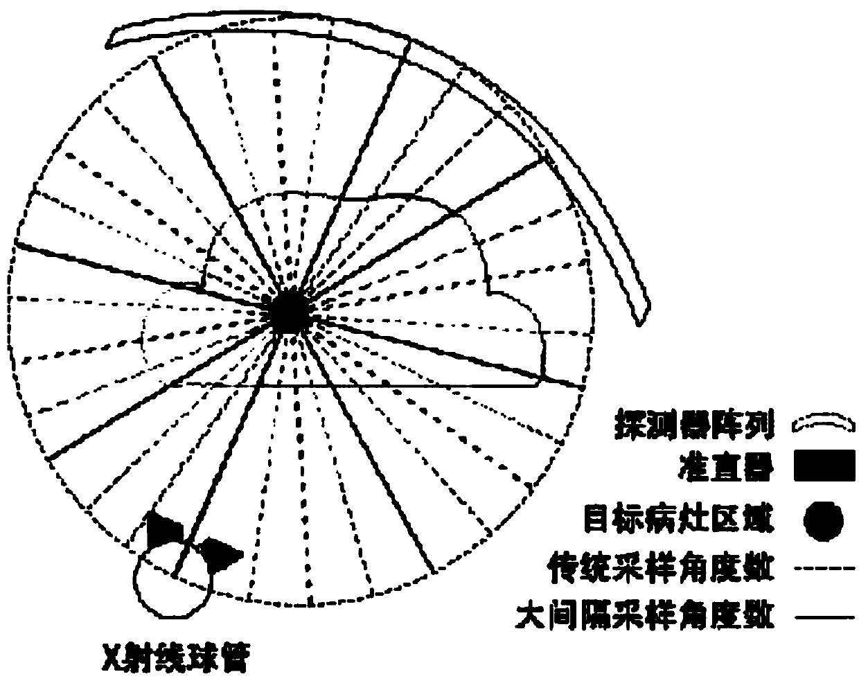
[0062] Step 2. Design the CT scanning method with limited number of projections. In this embodiment, the limited cross-sectional scanning range is related to the preset CT reconstruction range. Make the marker point at the rotation center of the CT scan, and select the detector width from the reconstruction range to ensure coverage of the entire reconstruction area; the design of the limi...


PUM


Abstract

The invention discloses a CT scanning method of low radiation dosage. A CT scanning manner of a limited X-ray projection beam is designed through the priori knowledge of the CT scanning of the previous focus of a patient, and through combination with the priori image of the CT scanning of the previous focus, the reconstruction of a CT image of an object region is performed. The CT scanning methodspecifically comprises the following steps of step 1, extracting a focus region in the priori CT scanning image in advance, positioning the object focus, and estimating the range required by CT scanning; step 2, according to different scanning requirements, designing the CT scanning manner and the scanning parameters of the limited X-ray projection beam so as to obtain the limited X-ray projectionbeam; and step 3, extracting the priori knowledge in the priori CT scanning image, performing adjustment so as to enable the priori CT scanning image to use the focus region as a center, and fusing the priori CT scanning image and the limited X-ray projection beam to reconstruct the CT image. Through the adoption of the CT scanning method disclosed by the invention, the X-ray radiation dosage received by human bodies in single-time CT scanning can be greatly reduced, the risk of inducing cancer can be reduced, and the requirements for reconstructing and detecting the focus region can be met.

Description

technical field [0001] The invention relates to the field of scanning and imaging of computed tomography (CT), in particular to a scanning method of computed tomography (CT) with low radiation dose. Background technique [0002] Computed tomography (CT) technology is more and more commonly used clinically because of its convenience in operation and effectiveness of detection. At the same time, the X-ray radiation brought by CT scanning has gradually been paid more attention. Excessive radiation doses can induce cancer. However, many clinical conditions require multiple CT scans in order to observe the lesions regularly. Therefore, reducing the radiation dose received by patients per CT scan has important clinical significance. One type of method is to reduce the intensity of X-rays to reduce the radiation dose, but the problem is that the noise signal of the CT image is relatively enhanced, and the image quality is degraded. Considering that many lesions are small in siz...

Claims


Application Information

Patent Timeline
IPC IPC(8): A61B6/03
Inventor 赵俊张伟康
Owner SHANGHAI JIAO TONG UNIV
Why Eureka
Try Eureka