Adsorption device, adsorption microscope detection device and laser scanning microscope
A detection device and microscope technology, which is applied in the field of laser scanning microscopes, can solve the problems of high resolution requirements for three-dimensional nonlinear laser scanning imaging, being easily affected by vibration, and affecting imaging quality, etc.
Pending Publication Date: 2019-05-14
BEIJING CHAOWEIJING BIOLOGICAL TECH CO LTD
0 Cites 0 Cited by
AI-Extracted Technical Summary
Problems solved by technology
[0004] However, the detection device based on the robotic arm in the above-mentioned 3D nonlinear laser scanning microscope has a large volume and the probe corresponds to a large skin area of the human body, so that the detection device is easily affected by ...
Method used
For the adsorption type three-dimensional nonlinear laser scanning microscope provided in each of the above-mentioned embodiments, the embodiments of the present invention also provide another specific implementation mode, and Fig. 14 provides the adsorption type three-dimensional nonlinear laser scanning microscope for the embodiments of the present invention A schematic diagram of a box-type combined structure, as shown in Figure 14, the scanning acquisition controller 531, industrial computer 532, air pumping device 52, fluorescence collecting device 56, femtosecond pulse laser and optical fiber coupling integration of the adsorption three-dimensional nonlinear laser scanning microscope The integrated modules 540 are integrated together in a portable suitcase, and there is an industrial computer with a display screen in the suitcase, and a display 55 is integrated on the cover of the suitcase; the adsorption microscope detection device 51 is adsorbed on the skin of the human body to be tested In terms of organization, it is connected with the optical fiber coupling in the box and the fluorescence collecting device 56 through optical fiber communication, connected with the suction pump through the suction pipeline, and the power plug is electrically connected with the scanning acquisition controller 531 and the industrial computer 532 . 15 is a schematic diagram of the box-sealing structure of the box-type combined structure of the adsorption-type three-dimensional nonlinear laser scanning microscope provided by the embodiment of the present invention. As shown in FIG. 15, the display 55 integrated on the box cover and the box body with various modules installed It is integrated together to facilitate the movement of the whole device and the replacement of the workplace, and the display 55 can be placed outside the cabinet when in use, so that the staff can obtain information on the display conveniently. After using the adsorption-type three-dimensional nonlinear laser scanning microscope, the staff can carry the equipment box to easily change the workplace, especially in hospitals, laboratories or outdoor places, it will be more convenient to use the equipment.
The adsorption type device that the embodiment of the present invention provides is used to arrange the miniature microscope probe and adopts outer casing, suction cup and cover glass to form the outer adsorption space that can make the adsorption device adsorb on the living body skin and is used to place the miniature microscope probe The inner closed space of the suction cup, wherein, the suction cup and the suction cup hole are connected by interference, so that the suction cup and the suction cup hole are in sealing contact, and the cover glass is fixed on the sealing port of the suction cup to realize the sealing contact, thereby forming an inner closed space and an outer adsorption space; the movement device as a whole It is fixed on the side wall of the outer casing, and the microscopic microscope probe moves up and down through the relative sliding of the limit block and the fixed bracket, and the microscopic microscope probe is aligned with the cover glass to output internal signals and signals through the cover glass. The external signal is received, and the overall device is miniaturized. After the device is adsorbed on the human skin, it can avoid the impact of life body activities on the microscopic microscope probe in the adsorption device. The operation is simple and easy to use.
The adsorption type three-dimensional non-linear laser scanning microscope that the embodiment of the present invention provides adopts fluorescence collection device...
Abstract
The embodiment of the invention provides an adsorption device, an adsorption microscope detection device and a laser scanning microscope. The adsorption device comprises a shell body, a sucking disc,a coverslip and a motion device, the sucking disc is embedded in a sucking disc hole, and the coverslip is fixed to a sealing opening of the sucking disc to form inner closed space and outer adsorption space; a fixed bracket of the motion device is fixed to the side wall of the shell body, and relative sliding connection between a limit block and the fixed bracket drives a minitype microscope probe to move up and down. According to the adsorption device, the adsorption microscope detection device and the laser scanning microscope, the outer adsorption space making the adsorption device adsorbed on the skin and the inner closed space used for arrangement of the probe are formed by the shell body, the sucking disc and the coverslip, so that after the adsorption device is adsorbed on the skin, the minitype microscope probe outputs an inner signal and receives an external signal by transmitting through the coverslip, the whole adsorption device is miniaturized, it is avoided that the probeis subjected to vibration influence caused by moving of a living body, and the adsorption device is simple in operation and convenient to use.
Application Domain
Diagnostic recording/measuringSensors
Technology Topic
PhysicsCover slip +3
Image
Examples
- Experimental program(1)
Example Embodiment
[0041] In order to make the purpose, technical solutions and advantages of the embodiments of the present invention clearer, the technical solutions in the embodiments of the present invention will be clearly and completely described below in conjunction with the drawings in the embodiments of the present invention. Obviously, the described embodiments It is a part of embodiments of the present invention, but not all embodiments. Based on the embodiments of the present invention, all other embodiments obtained by persons of ordinary skill in the art without creative efforts fall within the protection scope of the present invention.
[0042]The existing detection equipment based on fluorescence signals and second harmonic signals to obtain the morphology of biological cells is mainly a three-dimensional nonlinear laser scanning microscope. At present, the laser scanning microscope is mainly a laser scanning microscope based on a mechanical arm, namely The detection device of the laser scanning microscope is installed on the mechanical arm, and the detection device is moved through the adjustment of the mechanical arm, and then aligned to detect different tissue structures of the human body. However, due to the large volume of the laser scanning microscope, the probe corresponds to a large skin area of the human body, so that the detection device is easily affected by the vibration of the human body during specific operations, thereby affecting the imaging quality.
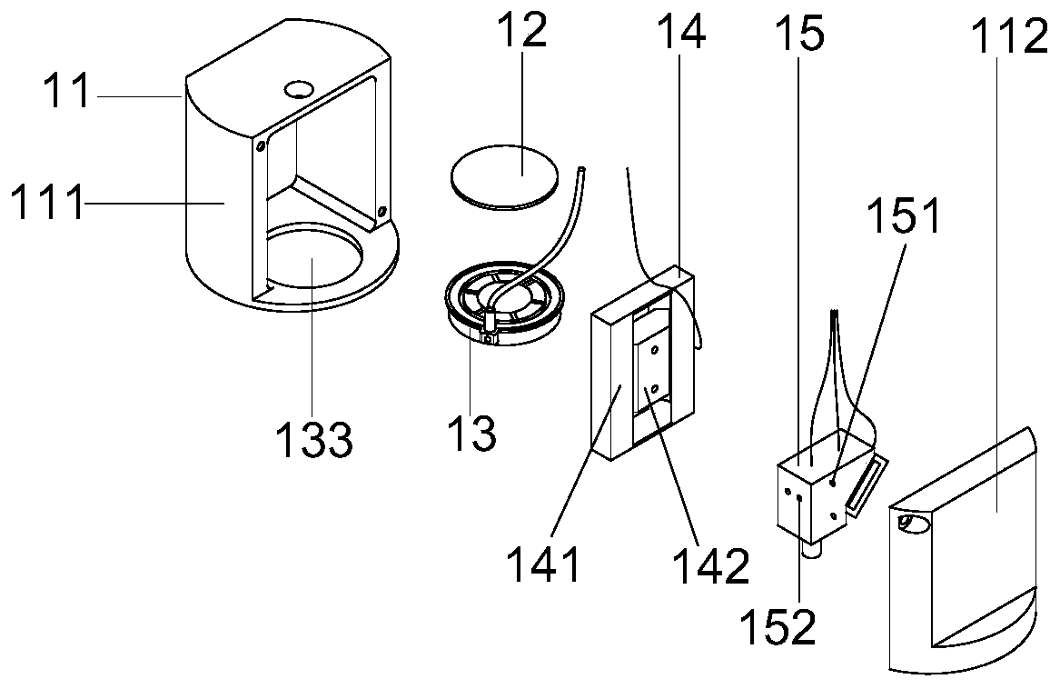
[0043] In order to more stably image the morphology of biological cells and obtain their structural information, an embodiment of the present invention provides an adsorption device for setting a microscopic microscope probe, figure 1 Schematic diagram of the structure of the adsorption device for setting the microscopic microscope probe provided by the embodiment of the present invention, such as figure 1 As shown, the adsorption device includes:
[0044] The outer casing 11, the suction cup 13, the cover glass 12 and the motion device 14 that drives the microscopic microscope probe 15 to move up and down, the suction cup 13 and the cover glass 12 are all arranged in the outer casing 11, wherein:
[0045] The suction cup 13 is embedded in the suction cup hole 133 opened at the bottom of the outer casing 11, the suction cup 13 is in interference connection with the suction cup hole 133, and the cover glass 12 is fixed on the sealing port of the suction cup 13 to form an inner closed space and an outer adsorption space of the adsorption device;
[0046] The moving device 14 is arranged in the inner closed space, and the moving device 14 includes a fixed bracket 141 and a limit block 142, wherein:
[0047] The fixed bracket 141 is fixed on the side wall of the outer shell 11 , and the limit block 142 is relatively slidably connected with the fixed bracket 141 to drive the microscopic microscope probe 15 to move up and down, wherein the microscopic microscope probe 15 is aligned with the cover glass 12 .
[0048] Specifically, the outer casing 11, the suction cup 13 and the cover glass 12 in the adsorption device for setting the microscopic microscope probe 15 provided by the embodiment of the present invention form an external adsorption space and an external adsorption space that enables the adsorption device to be adsorbed on the skin of a living body. In the inner closed space where the miniature microscope probe 15 is placed, the suction cup 13 and the cover glass 12 are at the bottom of the outer casing 11, and the bottom of the outer casing 11 is provided with a suction cup hole 133 for placing the suction cup 13, and the suction cup 13 and the suction cup hole 133 The interference connection makes the suction cup 13 and the suction cup hole 133 in sealing contact, and then the cover glass 12 is fixed on the sealing port of the suction cup 13 to realize the sealing contact, thereby forming an inner closed space and an outer suction space; the moving device 14 is integrally fixed on the outer casing 11 On the side wall, its limit block 142 slides up and down relative to the fixed bracket 141, the microscopic microscope probe 15 is fixed on the limit block 142, and under the drive of the limit block 142, it moves up and down, and the fixed bracket 141 and the micro microscope probe After the 15 is fixed, the microscopic microscope probe 15 is aligned with the cover glass 12 to output internal signals and receive external signals through the cover glass 12, so as to realize zooming and three-dimensional imaging of the microscope.
[0049] and the above figure 1 The outer casing 11, the cover glass 12, the sucker 13, and the moving device 14 are combined into a whole to be an adsorption device for setting the miniature microscope probe 15, and the miniature microscope probe 15 is fixed on the combined adsorption device to form a set. The adsorption device of the miniature microscope probe 15, wherein the miniature microscope probe 15 can be fixed on the limit block 142 of the moving device 14 by gluing, or can be detachably fixedly connected to the limit block 142 by bolt connection , that is, connect the moving device 14 and the microscopic microscope probe 15 through the bolt, the through hole on the limit block 141 and the first bolt hole 151 on the microscopic microscope probe 15, and also can increase between the limit block 141 and the microscopic microscope probe 15 Other fixing devices connect the moving device 14 and the micro microscope probe 15 through the through hole on the limit block and the second bolt hole 152 on the micro microscope probe 15 .
[0050] The adsorption device for setting the miniature microscope probe provided by the embodiment of the present invention adopts the outer shell, the suction cup and the cover glass to form the outer adsorption space that can make the adsorption device adsorb on the skin of the living body and the inner seal for placing the miniature microscope probe Space, wherein, the suction cup and the suction cup hole are connected by interference, so that the suction cup and the suction cup hole are in sealing contact, and the cover glass is fixed on the sealing port of the suction cup to achieve sealing contact, thereby forming an inner closed space and an outer adsorption space; the movement device is fixed on the outer shell as a whole On the side wall of the body, the micro-microscope probe moves up and down through the limit block and the fixed bracket, and the micro-microscope probe is aligned with the cover glass to output internal signals and receive external signals through the cover glass. , the overall device is miniaturized, and after the device is adsorbed on the human skin, it can avoid the impact of life body activities on the microscopic microscope probe in the adsorption device, and is easy to operate and use.
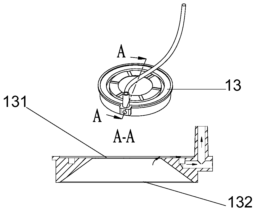
[0051] On the basis of the above-mentioned embodiments, the suction cup in the suction device for setting the microscopic microscope probe provided by the embodiment of the present invention also includes a suction port, figure 2 The schematic diagram of the sucker structure in the adsorption device for setting the microscopic microscope probe 15 provided by the embodiment of the present invention, as figure 2 As shown, the suction port 132 communicates with the sealing port 131, wherein:
[0052] The outer wall forming the suction opening 132 is in interference connection with the hole of the suction cup. That is, the suction cup 13 in the adsorption device provided in the embodiment of the present invention includes a suction port 132 in addition to the sealing port 131, and the sealing port 131 and the suction port 132 are connected, and the outer side wall of the suction port 132 is in interference connection with the suction cup hole, so that the suction cup It is in close contact with the outer shell, and then combined with a cover glass to form a sealed structure.
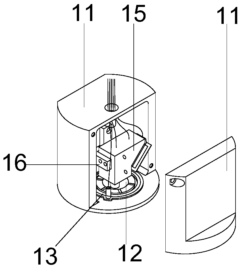
[0053] On the basis of the above-mentioned embodiments, the moving device in the adsorption device for setting the microscopic microscope probe provided by the embodiment of the present invention also includes a probe fixing frame, and the probe fixing frame is detachably fixedly connected with the limit block. The probe is detachably fixed on the limit block through the probe fixing frame. That is to say, the moving device in the adsorption device for setting the miniature microscope probe provided by the embodiment of the present invention also includes a probe fixing frame, wherein the probe fixing frame can be fixed on the limit block by screw connection, and the miniature microscope probe passes through the probe The fixed frame is fixed on the limit block, wherein, image 3 The schematic diagram of the combined structure of the adsorption device for setting the microscopic microscope probe provided by the embodiment of the present invention, as shown in image 3 As shown, the suction cup 13 is embedded in the suction cup hole of the outer casing 11, the cover glass 12 is fixed on the suction cup 13, and the microscopic microscope probe 15 is fixed on the limit block through the probe holder 16, that is, through the through hole on the limit block and figure 1 The second bolt hole 152 shown fixes the microscopic microscope probe 15 on one side of the probe fixing frame 16 , and fixes the stopper on the other side, and makes the microscopic microscope probe 15 align with the cover glass 12 at the same time. in, Figure 4 The schematic diagram of the structure of the probe holder in the adsorption device for setting the microscopic microscope probe provided by the embodiment of the present invention, as shown in Figure 4 As shown, the probe fixing frame is provided with a limit block through hole 161 and a probe through hole 162, the limit block through hole 161 is used for connecting a fixed limit block, and the probe through hole 162 is used for connecting a microscopic microscope probe.
[0054] in, Figure 5 The schematic diagram of the cross-sectional structure of the combined adsorption device for setting the microscopic microscope probe provided by the embodiment of the present invention, such as Figure 5 As shown, the suction cup 13 is embedded in the suction cup hole of the outer shell 11, the cover glass 12 is fixed on the suction cup 13, the microscopic microscope probe 15 is fixed on the limit block, and the microscopic microscope probe 15 is positively aligned with the cover glass 12, and The microscopic microscope probe is located in the inner closed space 17, and the adsorption device is adsorbed on the skin and other tissues of the living body to be tested by the negative pressure formed in the outer adsorption space 18.
[0055] On the basis of the above-mentioned embodiments, the adsorption device for setting the microscopic microscope probe provided by the embodiment of the present invention also includes a motor, and the motor is arranged in the inner closed space, wherein:
[0056] The motor is rotationally connected with the limit block through a lead screw, and is used to drive the limit block to move up and down. That is to say, the adsorption device for setting the miniature microscope probe provided by the embodiment of the present invention is also provided with a motor, which is used to drive the stopper with the miniature microscope probe to move up and down, so as to adjust the external focal length of the miniature microscope probe, thereby realizing Three-dimensional detection at different depths and levels.
[0057] On the basis of the above-mentioned embodiments, the outer casing in the adsorption device for setting the microscopic microscope probe provided by the embodiment of the present invention includes a first casing and a second casing, such as figure 1 shown, where:
[0058] An accommodating space is provided in the first housing 111 , the suction cup and the cover glass are both disposed in the accommodating space, and the first housing 111 and the second housing 112 are detachably fixedly connected. That is to say, the outer casing in the adsorption device for setting the microscopic microscope probe provided by the embodiment of the present invention includes two parts, which are respectively the first casing 111 and the second casing 112, and the first casing 111 is used for setting the suction cup and the cover. The accommodating space for the glass slide, that is, the suction cup hole in the above embodiment is also arranged at the bottom of the first housing 111, and the suction cup is embedded in the suction cup hole of the first housing 111, and combined with the cover glass and the second housing 112, it can be The outer suction space and the inner closed space in the above embodiment are formed, wherein the first shell 111 and the second shell 112 can be detachably fixedly connected by screws, which facilitates the assembly and disassembly of the whole device and the replacement of parts.
[0059] An embodiment of the present invention also provides an adsorption-type microscope detection device, which includes a miniature microscope probe and the adsorption device for setting the miniature microscope probe provided in the above-mentioned embodiments, wherein the miniature microscope probe is detachable Fixed on the limit block, the miniature microscope probe includes a probe housing and a fixed plate, the top of the probe housing is provided with a first port and a second port, and the bottom of the probe housing is provided with a third port, wherein:
[0060] A first optical path is provided in the first channel formed between the first port and the third port, and a second optical path is provided in the second channel formed between the second port and the third port, wherein:
[0061] The first optical path sequentially includes a collimator lens, a microelectromechanical scanning galvanometer, a lens, a dichroic mirror, and an objective lens between the first port and the third port, wherein the first optical path is used to conduct the light received by the first port. The laser signal is from the first port to the third port;
[0062] The second optical path sequentially includes an objective lens and a dichroic mirror located between the third port and the second port, wherein the second optical path is used to transmit the optical signal collected by the objective lens from the third port to the second port.
[0063] specifically, Image 6 Schematic diagram of the structure of the miniature microscope probe in the adsorption microscope detection device provided by the embodiment of the present invention Figure 1 ,like Image 6As shown, the probe housing 150 of the above-mentioned miniature microscope probe is provided with three ports, two channels are formed between the three ports, and two optical paths are arranged in the two channels, which are respectively the first optical path and the second optical path. Optical path, there is an overlapping part between the two channels formed between the three ports, and the dichroic mirror 1544 and objective lens 1545 shared by the above two optical paths are arranged in the overlapping part of the channel, wherein the first optical path is The emitting light path includes in turn a collimating lens 157 between the first port 154 and the third port 156, a micro-electromechanical scanning vibrating mirror 1541, a lens 1542, a dichroic mirror 1543 and an objective lens 1544, and the objective lens is fixed by a fixing frame 159 On the probe housing 150, that is, the laser signal enters from the first port 154, conducts through the first optical path, and exits from the third port 156, and the parallel light incident on the first port passes through two lenses, and then The way of parallel light is emitted; the second optical path is a collection optical path, which is used to output the collected optical signal from the second port 155 to the collection device connected with the microscopic microscope probe; and the laser signal transmitted by the first optical path is transmitted in two directions Under the reflection of the color mirror 1543, it can be vertically and positively incident into the objective lens 1544. The dichroic mirror reflects the laser signal of the first optical path and transmits the optical signal collected by the second optical path, and the pitch angle of the dichroic mirror 1543 can be adjust.
[0064] On the basis of the above-mentioned embodiments, the adsorption microscope detection device provided by the embodiment of the present invention also includes:
[0065] A liquid lens, the liquid lens is located between the collimating lens and the micro-electromechanical scanning galvanometer to form a new first optical path, and the new first optical path sequentially includes a collimating lens located between the first port and the third port, Liquid lens, MEMS scanning mirror, lens, dichroic mirror and objective lens. That is, a liquid lens 158 can be added to the first optical path of the miniature microscope probe in the adsorption microscope detection device provided by the embodiment of the present invention, Figure 7 Schematic diagram of the structure of the miniature microscope probe in the adsorption microscope detection device provided by the embodiment of the present invention Figure II ,like Figure 7 As shown, through the Figure 7 A liquid lens 158 is added to the probe structure of the miniature microscope shown to realize the zoom function. The liquid lens 158 is positioned between the collimator lens 157 and the microelectromechanical scanning vibrating mirror 1541, at Figure 7 In the shown original optical path structure, the laser signal becomes parallel light after passing through the collimating lens 157 . After the liquid lens 158 is added, the surface of the liquid lens 158 is curved accordingly by applying a voltage or current to the liquid lens 158, thereby producing different optical powers for parallel light. The specific optical path is as follows: the laser signal exits from the optical fiber, passes through the collimator lens 157, and then enters the liquid lens 158 in parallel. The corresponding optical power is generated from the liquid lens 158 according to the applied voltage or current signal, and the outgoing converging or diverging light passes through the micro-electromechanical After scanning the galvanometer 1541, the lens 1542, the dichroic mirror 1543 and the objective lens 1544, they converge onto the sample. Among them, the focal power change introduced by the liquid lens 158 will cause the focal point of the laser signal emitted from the objective lens 1544 to move back and forth in the depth direction, and the response speed of the liquid lens 158 is very fast, and its scanning frequency is on the order of KHz, so it can realize Fast depth-wise scanning imaging. Wherein, the liquid lens 158 is equivalent to parallel flat glass when no voltage or current signal is applied, and has no optical power for the laser signal and does not cause any shift of the focal point behind the objective lens. In specific use, the liquid lens 158 is complementary to the moving device, and the position of the objective lens 1544 is adjusted through the moving device. After roughly adjusting to the corresponding depth position, the system switches to the zoom scanning mode of the liquid lens 158 to perform fast three-dimensional imaging on the sample. When the adsorption microscope detection device is not equipped with a moving device, the zoom adjustment can also be performed only through the liquid lens.
[0066] The embodiment of the present invention also provides an adsorption three-dimensional nonlinear laser scanning microscope, Figure 8 Schematic diagram of the structure of the adsorption-type three-dimensional nonlinear laser scanning microscope provided for the embodiment of the present invention, such as Figure 8 As shown, the laser scanning microscope includes:
[0067] Fluorescence collection device 56, pumping device 52, scanning acquisition controller 531, femtosecond pulse laser 541, fiber coupling module 542, and the adsorption microscope detection device 51 provided in the above embodiment, fluorescence collection device 56 and fiber coupling module 542 are all compatible with The adsorption microscope detection device 51 is connected by optical fiber communication, the fluorescence collection device 56 and the adsorption microscope detection device 51 are electrically connected to the scanning acquisition controller 531, and the air extraction device 52 is electrically connected to the adsorption microscope detection device 51, wherein:
[0068] A femtosecond pulsed laser 541, configured to output a pulsed laser signal to a fiber coupling module 542;
[0069] The fiber coupling module 542 is used to couple the pulsed laser signal output by the femtosecond pulsed laser 541, and transmit the pulsed laser signal to the miniature microscope probe in the adsorption microscope detection device;
[0070] The adsorption microscope detection device is used to receive the pulsed laser signal, output the pulsed laser signal to the autofluorescence substance in the living body cells, obtain the fluorescence signal and the second harmonic signal generated by the excitation of the autofluorescence substance, and output the fluorescence signal and the second harmonic signal to the fluorescence collecting device 56;
[0071] The fluorescence collecting device 56 is used to convert the fluorescence signal and the second harmonic signal into corresponding electrical signals after receiving the fluorescence signal and the second harmonic signal;
[0072] Scanning acquisition controller 531, used to control the microscopic microscope probe to scan the pulsed laser signal, and synchronously acquire electrical signals;
[0073] The air extraction device 52 is used to extract air from the outer adsorption space of the adsorption microscope detection device, so as to form a negative pressure in the outer adsorption space.
[0074] Specifically, the adsorption three-dimensional nonlinear laser scanning microscope provided by the embodiment of the present invention includes a fluorescence collection device 56, an air extraction device 52, a scanning acquisition controller 531, a femtosecond pulse laser 541, a fiber coupling module 542, and an adsorption microscope detection device 51, so as to form a three-dimensional nonlinear laser scanning microscope that can be adsorbed on the human skin or penetrate into the human stomach for detection, wherein the femtosecond pulsed laser 541 can emit pulsed laser signals to excite the autofluorescent substances in human skin cells to generate multiphotons Fluorescence signal and second harmonic signal, including using 920nm femtosecond pulsed laser 541 to excite FAD and collagen in cells, exciting 500-600nm fluorescence signal and 460nm second harmonic signal, and passing 780nm femtosecond pulse The laser 541 excites autofluorescent substances such as FAD or NADH in the cells to generate corresponding fluorescent signals and second harmonic signals;
[0075] Among them, the fluorescence collection device 56 integrates two signal collection optical paths, which are respectively the fluorescence signal collection optical path and the second harmonic signal collection optical path, to realize the separate collection of the fluorescence signal and the second harmonic signal; the scanning acquisition controller 531 controls the micro The scanning galvanometer in the microscope probe scans the pulsed laser signal and excites the autofluorescent substance to generate a fluorescent signal and a second harmonic signal, and collects the first electrical signal and The second electrical signal; the air extraction device 52 mainly includes an air extraction pump, which is connected with the air extraction pipeline, and an air extraction valve is arranged in the air extraction pipeline, and the air extraction valve is electrically connected with the air extraction device 52, and the air extraction device 52 is adjusted by adjusting the air extraction valve. The switch and the size of the opening and closing control the suction flow of the extraction pipeline, so as to realize the control of the suction of the external adsorption space, and then adjust the negative pressure in the external adsorption space, so that the adsorption device can be adsorbed on the skin of the living body through the effect of atmospheric pressure. On tissues such as the stomach and intestines, and the adsorption-type three-dimensional nonlinear laser scanning microscope can include two-photon scanning microscopes and multi-photon scanning microscopes according to classification. Among them, when femtosecond pulsed lasers can be replaced by ordinary continuous light lasers, increase The pinhole diaphragm can also be adjusted for the adsorption-type 3D nonlinear laser scanning microscope as a confocal microscope. Among them, the resolution of the adsorption three-dimensional nonlinear laser scanning microscope can be set to 800nm, the imaging field of view can be 200 microns*200 microns, and the imaging speed can be 26 frames (256*256 pixels) or 13 frames (512*512 pixels) .
[0076] The adsorption-type three-dimensional nonlinear laser scanning microscope provided by the embodiment of the present invention adopts a fluorescence collection device, an air extraction device, a scanning acquisition controller, a femtosecond pulse laser, an optical fiber coupling module, and an adsorption-type microscope detection device, thereby forming a microscope that can be adsorbed on human skin. Or a three-dimensional nonlinear laser scanning microscope that penetrates into the human stomach and intestines. By adjusting the distance between the microscopic microscope probe and the cover glass to adjust the focal length, the three-dimensional scanning of the laser scanning microscope can be realized. The femtosecond pulse laser excites intracellular autofluorescent substances to obtain multiple Photonic fluorescence signals and second harmonic signals realize the nonlinearity of the laser scanning microscope. The fluorescence signals and second harmonic signals are collected by the fluorescence collection device and converted into corresponding electrical signals, and then the corresponding reflected cells are obtained through the electrical signals. Fluorescence images of tissue structures, etc. Among them, the use of adsorption microscope detection devices can avoid the impact of life body activities on the vibration of the micro-microscope probe, thereby avoiding the impact of vibration on the imaging quality, and the above-mentioned laser scanning microscope can realize multiple imaging modes including XY Imaging, XZ imaging and 3D imaging, where XY imaging is horizontal scanning imaging at a certain depth of the cell structure, XZ imaging is XZ section imaging from the surface to a certain depth, 3D imaging is from the surface to a certain depth, XY imaging is performed at each depth and reconstructed into a 3D image. The equipment is easy to operate and easy to use.
[0077] On the basis of above-mentioned each embodiment, Figure 9 Schematic diagram of the structure of the fluorescence collecting device provided for the embodiment of the present invention, such as Figure 9 As shown, the fluorescence collection device provided by the embodiment of the present invention includes an optical fiber universal interface 781, a first photomultiplier tube 782, a second photomultiplier tube 783, and a first collector located between the optical fiber universal interface 781 and the first photomultiplier tube 782. Optical path, the second collection optical path between the optical fiber universal interface 781 and the second photomultiplier tube 783, wherein:
[0078] The first collection optical path includes a coupling collection lens 71, an infrared filter 72, a first dichroic mirror 73, a first optical filter 74, and a first collection lens 75 in sequence, wherein the first collection optical path is used to collect the fluorescence collection device For the received fluorescent signal, the first photomultiplier tube 782 is used to convert the fluorescent signal into a first electrical signal;
[0079] The second collection optical path includes a coupling collection lens 71, an infrared filter 72, a first dichroic mirror 73, a second dichroic mirror 76, a second filter 77 and a second collection lens 78 in turn, wherein the second The collection optical path is used to collect the second harmonic signal received by the fluorescence collection device, and the second photomultiplier tube 783 is used to convert the second harmonic signal into a second electrical signal. That is, the fluorescence collection device provided by the embodiment of the present invention has a dual-channel signal collection function and integrates two optical paths, wherein the first dichroic mirror 73 in the first collection optical path transmits the fluorescent signal and reflects the second harmonic dichroic mirror 73. The dichroic mirror, the second dichroic mirror 76 and the first dichroic mirror 73 are the same dichroic mirror for reflecting the second harmonic, and the first optical filter 74 is used for transmitting the fluorescent signal and filtering out the rest interference signal, the second optical filter 77 is used to transmit the corresponding second harmonic signal to filter out the rest of the interference signal, for example, when using a 780nm femtosecond fiber laser to excite the autofluorescent substances in the skin cells on the surface of the human body, a 390nm The second harmonic signal and the two-photon autofluorescence signal of 450-600nm pass through the wavelength above 420nm, and the dichroic mirror reflected by the wavelength below 420, that is, the first dichroic mirror 73 can separate the two fluorescence channels, respectively using 390±20nm The first optical filter 74 and the second optical filter 77 of 450-600nm can obtain clean second harmonic signal and fluorescence signal.
[0080] On the basis of the above-mentioned embodiments, the adsorption-type three-dimensional nonlinear laser scanning microscope provided by the embodiment of the present invention also includes an industrial computer, such as Figure 8 As shown, the industrial computer 532 is electrically connected to the scanning acquisition controller 531, wherein:
[0081]The industrial computer 532 is used to acquire the first electrical signal and the second electrical signal collected by the scanning acquisition controller 531 , and generate a first fluorescence image based on the first electrical signal and a second fluorescence image based on the second electrical signal. That is, the adsorption-type three-dimensional nonlinear laser scanning microscope provided in the embodiment of the present invention further includes an industrial computer 532 electrically connected to the scanning acquisition controller 531, and the industrial computer 532 generates the first fluorescence image based on the first electrical signal and generates the first fluorescence image based on the second electrical signal. Generate the second fluorescence image, which can be used to display the cell structure and fiber structure information respectively, wherein the control software is installed on the industrial computer, and through the control software, control instructions are sent to the scanner to control the scanning acquisition controller to obtain the above-mentioned first electric signal and a second electrical signal.
[0082] On the basis of the above-mentioned embodiments, the adsorption-type three-dimensional nonlinear laser scanning microscope provided by the embodiment of the present invention also includes a display, such as Figure 8 As shown, the display 55 is electrically connected to the industrial computer 532 for displaying the first fluorescent image and the second fluorescent image. That is, the adsorption-type three-dimensional nonlinear laser scanning microscope provided in the embodiment of the present invention also includes a display 55 for displaying the first fluorescence image and the second fluorescence image. Through the display 55, the staff can directly obtain the first fluorescence image and the second fluorescence image. Information about the image.
[0083] On the basis of the foregoing embodiments, there are multiple adsorption microscope detection devices in the adsorption three-dimensional nonlinear laser scanning microscope provided in the embodiments of the present invention. That is, the fluorescence collecting device and the fiber coupling module provided by the embodiment of the present invention can be simultaneously connected with multiple adsorption microscope detection devices through optical fiber communication, that is, multiple detection devices are integrated in an adsorption three-dimensional nonlinear laser scanning microscope system to realize the detection of Simultaneous detection of different tissue parts of the living body for comparative analysis.
[0084] On the basis of the above-mentioned embodiments, the adsorption-type three-dimensional nonlinear laser scanning microscope provided by the embodiment of the present invention also includes an adjustment optical fiber, which is used for the optical fiber transmission between the fluorescence collection device and the fiber coupling module and the adsorption-type microscope detection device respectively. connection, where:
[0085] The length of the adjustment fiber is adjustable. That is, the fluorescence collecting device and the fiber coupling module in the adsorption-type three-dimensional nonlinear laser scanning microscope provided by the embodiment of the present invention are respectively connected to the adsorption-type microscope detection device through an adjustable optical fiber with an adjustable length, so as to realize the requirements of different experimental scenarios. , to flexibly move the detection device to avoid the limitation of the limited fiber length, wherein the length of the adjusting fiber can be adjusted, in order to realize the application of various occasions by replacing the fiber of different lengths, the fiber of different lengths can be replaced at any time according to the needs.
[0086] In order to more clearly illustrate the application scenarios of the adsorption-type three-dimensional nonlinear laser scanning microscope provided by the embodiment of the present invention, it is further described with illustrations, Figure 10 The schematic diagram of the adsorption-type three-dimensional nonlinear laser scanning microscope for detecting human facial skin tissue provided by the embodiment of the present invention, as shown in 10, through the suction function of the suction device 52, the adsorption-type microscope detection device 51 is adsorbed on the human face , wherein, the first device 53 integrates a scanning acquisition controller and an industrial computer, the industrial computer is electrically connected to the display 55, and the second device 54 integrates a femtosecond pulse laser, a fiber coupling module and a fluorescence collection device, and the fiber coupling module and fluorescence The collection devices are all connected to the adsorption microscope detection device 51 through optical fiber transmission, wherein the working principle of the adsorption three-dimensional nonlinear laser scanning microscope is the same as that of the above-mentioned embodiments, and will not be repeated here.
[0087] in, Figure 11 The schematic diagram of the adsorption-type three-dimensional nonlinear laser scanning microscope detection of human chest skin tissue provided by the embodiment of the present invention, as shown in 11, the adsorption-type microscope detection device 51 is adsorbed on the human chest through the pumping function of the air pumping device 52, wherein , the first device 53 integrates a scanning acquisition controller and an industrial computer, the industrial computer is electrically connected to the display 55, and the second device 54 integrates a femtosecond pulse laser, a fiber coupling module and a fluorescence collection device, and the fiber coupling module and the fluorescence collection device They are all connected to the adsorption microscope detection device 51 through optical fiber transmission, wherein the working principle of the adsorption three-dimensional nonlinear laser scanning microscope is the same as that of the above-mentioned embodiments, and will not be repeated here.
[0088] in, Figure 12 A schematic diagram of multiple detection devices of an adsorption-type three-dimensional nonlinear laser scanning microscope provided for an embodiment of the present invention to simultaneously detect human skin tissue, as shown in 12, through the pumping function of the air pumping device 52, a plurality of adsorption-type microscope detection devices 51 They are adsorbed on the face, chest and legs of the human body at the same time, wherein the first device 53 integrates a scanning acquisition controller and an industrial computer, and the industrial computer is electrically connected to the display 55, and the second device 54 integrates a femtosecond pulse laser, an optical fiber The coupling module and the fluorescence collection device, the optical fiber coupling module and the fluorescence collection device are all connected to the adsorption microscope detection device 51 through optical fiber transmission, so that under the action of multiple adsorption microscope detection devices, the skin tissue structure of different parts of the human body can be detected simultaneously. The operation is simple and easy to use, and since the optical fiber communication connection is adopted between the adsorption microscope detection device and the second device, the length of the optical fiber can be adjusted, so that the human body to be tested can move freely. Wherein, the working principle of the adsorption-type three-dimensional nonlinear laser scanning microscope is the same as that of the above-mentioned embodiments, and will not be repeated here. Figure 13 Schematic diagram of the adsorption-type three-dimensional nonlinear laser scanning microscope for detecting animal skin tissue provided by the embodiment of the present invention, as shown in Figure 13 As shown, the adsorption microscope detection device 51 can also be adsorbed on the skin tissue of the living body through the air pumping function of the air pumping device 52, wherein the first device 53 integrates a scanning acquisition controller and an industrial computer, and the industrial computer It is electrically connected with the display 55, and the second device 54 integrates a femtosecond pulsed laser, a fiber coupling module and a fluorescence collecting device. The embodiment is the same.
[0089] For the adsorption-type three-dimensional nonlinear laser scanning microscope provided in the above-mentioned embodiments, the embodiment of the present invention also provides another specific implementation mode, Figure 14 A schematic diagram of a box-type combined structure of an adsorption-type three-dimensional nonlinear laser scanning microscope is provided for an embodiment of the present invention, as shown in Figure 14 As shown, the scanning acquisition controller 531, industrial computer 532, air pumping device 52, fluorescence collecting device 56, femtosecond pulsed laser and optical fiber coupling integrated module 540 of the adsorption-type three-dimensional nonlinear laser scanning microscope are integrated together In the portable suitcase, there is an industrial computer with a display screen in the suitcase, and a display 55 is integrated on the cover of the suitcase; the adsorption microscope detection device 51 is adsorbed on the skin tissue of the human body to be tested, and coupled with the optical fiber in the suitcase. It is connected with the fluorescence collecting device 56 through optical fiber communication, connected with the suction pump through the suction pipeline, and the power plug is electrically connected with the scanning acquisition controller 531 and the industrial computer 532 . in Figure 15 A schematic diagram of the box-sealing structure of the box-type combined structure of the adsorption-type three-dimensional nonlinear laser scanning microscope provided in the embodiment of the present invention, as shown in Figure 15 As shown, the display 55 integrated on the box cover is integrated with the box body installed with various modules, which facilitates the movement of the whole device and the replacement of the workplace, and the display 55 can be placed on the box body when in use for convenience. Workers access the information on the display. After using the adsorption-type three-dimensional nonlinear laser scanning microscope, the staff can carry the equipment box to easily change the workplace, especially in hospitals, laboratories or outdoor places, it will be more convenient to use the equipment.
[0090] It should be further explained that, the adsorption-type three-dimensional nonlinear laser scanning microscope provided by the above-mentioned embodiments, after changing the wavelength of the femtosecond pulse laser and adjusting the filter range of each filter, the partial fluorescence and non-SHG (SecondHarmonic Generation , second harmonic generation) signal active tissue, the CARS signal can be collected, and thus adjusted to an adsorption micro-CARS microscope, and the specific adjustment parameters can be set according to specific needs.
[0091] Although the specific implementation of the present invention has been described above in conjunction with the accompanying drawings, it does not limit the scope of protection of the invention. Those skilled in the art should understand that on the basis of the technical solution of the present invention, those skilled in the art do not need the above only The preferred embodiments of the present application are not intended to limit the present invention. For those skilled in the art, there may be various modifications and changes in the present application. Any modifications, equivalent replacements, improvements, etc. made within the spirit and principles of this application shall be included within the protection scope of this application.
[0092] The device embodiments described above are only illustrative, and the units described as separate components may or may not be physically separated, and the components shown as units may or may not be physical units, that is, they may be located in one place , or can also be distributed to multiple network elements. Part or all of the modules can be selected according to actual needs to achieve the purpose of the solution of this embodiment. It can be understood and implemented by those skilled in the art without any creative efforts.
[0093] Finally, it should be noted that: the above embodiments are only used to illustrate the technical solutions of the present invention, rather than to limit them; although the present invention has been described in detail with reference to the foregoing embodiments, those of ordinary skill in the art should understand that: it can still be Modifications are made to the technical solutions described in the foregoing embodiments, or equivalent replacements are made to some of the technical features; and these modifications or replacements do not make the essence of the corresponding technical solutions deviate from the spirit and scope of the technical solutions of the various embodiments of the present invention.
PUM


Description & Claims & Application Information
We can also present the details of the Description, Claims and Application information to help users get a comprehensive understanding of the technical details of the patent, such as background art, summary of invention, brief description of drawings, description of embodiments, and other original content. On the other hand, users can also determine the specific scope of protection of the technology through the list of claims; as well as understand the changes in the life cycle of the technology with the presentation of the patent timeline. Login to view more.