Method, system and computing device for automatic detection of infant hip joint angle

An automatic detection and hip joint technology, applied in the field of medical ultrasound detection, can solve the problems of long-term identification and long time to obtain results, etc.

Active Publication Date: 2020-11-10
GUANGDONG WOMEN & CHILDREN HOSPITAL
View PDF5 Cites 0 Cited by
  • Summary
  • Abstract
  • Description
  • Claims
  • Application Information

AI Technical Summary

Problems solved by technology

This evaluation method has many marked points on the ultrasound image, and it takes a long time for the sonographer to identify and obtain standard section measurements during the examination; after obtaining the image data, the sonographer has to use another instrument for measuring angles, and the hip joint needs to be measured twice. The data of two angles can be obtained only after the operation, and it takes a long time to obtain the result

Method used


Image

  • Method, system and computing device for automatic detection of infant hip joint angle
  • Method, system and computing device for automatic detection of infant hip joint angle
  • Method, system and computing device for automatic detection of infant hip joint angle

Examples


Embodiment 1

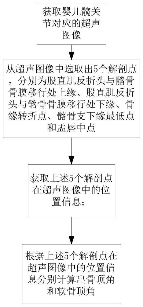
[0069] This embodiment discloses a method for automatically detecting the angle of an infant's hip joint, such as figure 1 As shown, the steps are as follows:

[0070] Step S1. Obtain the ultrasonic image corresponding to the baby's hip joint; in this embodiment, the obtained ultrasonic image is a preprocessed image, wherein the preprocessing includes image noise reduction and size processing. In this embodiment, the preprocessed The ultrasonic image obtained after processing can be an image with a size of 1280*872.

[0071] Step S2: Select 5 anatomical points from the ultrasound image, which are the upper edge of the rectus femoris reflex head and the transition of the iliac periosteum, the lower edge of the rectus femoris reflex head and the ilium periosteum transition, the bone edge turning point, The lowest point of the lower edge of the iliac ramus and the midpoint of the labrum;

[0072] When selecting 5 anatomical points from the ultrasound image in this embodiment, t...

Embodiment 2

[0093] This embodiment discloses an automatic detection system for infant hip joint angles, such as figure 2 As shown, it includes an ultrasonic image acquisition module, an anatomical point selection module, an anatomical point position acquisition module and a hip joint angle calculation module, wherein the specific functions of the above-mentioned modules are as follows:

[0094] The ultrasonic image acquisition module is used to obtain the corresponding ultrasonic image of the infant's hip joint; based on this module, the user can import the ultrasonic image that needs to be tested for the infant's hip joint angle into the system.

[0095] The anatomical point selection module is used to select 5 anatomical points from the ultrasound image, which are the upper edge of the rectus femoris reflex head and the transition of the ilium periosteum, the lower edge of the rectus femoris reflex head and the ilium periosteum transition, The turning point of the bony margin, the lowe...

Embodiment 3

[0119] This embodiment discloses a computing device, including a processor and a memory for storing a program executable by the processor. When the processor executes the program stored in the memory, the following operations are realized:

[0120] Obtain the ultrasonic image corresponding to the baby's hip joint; in this embodiment, the obtained ultrasonic image is a preprocessed image, wherein the preprocessing includes image noise reduction processing and size processing, and in this embodiment, after preprocessing, the obtained The ultrasound image can be a 1280*872 image.

[0121] Five anatomical points were selected from the ultrasonic images, namely, the upper edge of the rectus femoris reflex head and the transition of the ilium periosteum, the lower edge of the rectus femoris muscle and the transition of the ilium periosteum, the turning point of the bone border, and the ilium ramus. The lowest point of the lower edge and the midpoint of the labrum; in this embodiment...


PUM


Abstract

The invention discloses a Infant hip joint angle automatic detection method and system and a computing device. The method comprises the following steps of firstly, ultrasonic images corresponding to hip joints of infants are obtained, and five dissection points are selected from the ultrasonic images and are the upper edge of the femoral rectus retroflexion head and iliac periosteum moving position, the lower edge of the femoral rectus retroflexion head and iliac periosteum moving position, the bone edge retroflexion point, the lowest point of the iliac branch lower edge and the pelvis midpoint; position information of each anatomical point is acquired in the ultrasonic image; a bone vertex angle alfa and a cartilage vertex angle Beta are respectively calculated according to the position information of each anatomical point in the ultrasonic image; in the method, the automatic measurement of the hip joint angle can be completed only by selecting five anatomical points from the ultrasonic image by an ultrasonic doctor. The technical problems that in the prior art, a straight line needs to be drawn on an ultrasonic image manually, the cartilage vertex angle is measured through an instrument, the measurement time is long, operation is troublesome, and errors are prone to occurring are solved, and the advantages of high baby hip joint angle detection speed and high accuracy are achieved.

Description

technical field [0001] The invention relates to the technical field of medical ultrasonic detection, in particular to an automatic detection method, system and computing equipment for an infant's hip joint angle. Background technique [0002] The hip joint connects the trunk and the lower limbs. It is the most needed and critical part in walking and sports. In medical research, it is often necessary to use ultrasound to observe and analyze the baby's hip joint, especially when the femoral head is not fully developed. Angle measurement and analysis of the hip joint during ossification. [0003] The Graf examination method is an ultrasonic method commonly used in the world and in China to measure the hip joint in infants under static conditions. By observing the shape of the acetabulum, the positional relationship between the femoral head and the acetabulum, and measuring the data of the two angles of the joint, the hip joint can be divided into two types: Four types and nine...

Claims


Application Information

Patent Timeline
Patent Type & Authority Patents(China)
IPC IPC(8): G06T7/00
Inventor 陈丹陈涛唐遥芳陈敏莹谭蕴宜
Owner GUANGDONG WOMEN & CHILDREN HOSPITAL
Why Eureka
Try Eureka