Gene heterogeneity visual quantification method in glioma based on pyradiomics and system

A radiomics and glioma technology, applied in the field of medical and radiomics, can solve the problems of bleeding and trauma, lack of research, delineate gene heterogeneity map, etc., achieve accurate radiotherapy and chemotherapy sensitivity, significant clinical value, Improved Survival Outcomes

Active Publication Date: 2019-08-06
FUDAN UNIV
View PDF7 Cites 6 Cited by
  • Summary
  • Abstract
  • Description
  • Claims
  • Application Information

AI Technical Summary

Problems solved by technology

[0005]Currently, the research bottleneck for glioma heterogeneity lies in two aspects: the research method is relatively complicated, especially for the study of in vivo tumors, it must be based on MRI navigation Point puncture sampling, so that tissue samples can be accurately obtained, and more accurate gene sequencing can be performed. However, multi-point puncture takes a long time, and the most critical risk is bleeding and trauma; most of them are "point-to-point" research and lack "face-to-face" research. Research
The curr

Method used


Image

  • Gene heterogeneity visual quantification method in glioma based on pyradiomics and system
  • Gene heterogeneity visual quantification method in glioma based on pyradiomics and system
  • Gene heterogeneity visual quantification method in glioma based on pyradiomics and system

Examples


Embodiment Construction

[0029] The radiomics-based visualization and quantification method for gene heterogeneity in glioma of the present invention will be further described below with a specific example of IDH1.

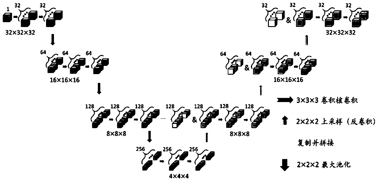
[0030] Step 1 is image segmentation. The network structure adopted in the present invention is as figure 1 As shown, the downsampling path and the upsampling path are combined in series with the feature maps of the corresponding resolution levels. The downsampling path has three blocks and two convolutional layers. Each block consists of two consecutive convolutional layers with a filter size of 3×3×3 and a max-pooling layer with a stride of 2×2×2. At the end of the downsampling path, two successive convolutional layers are added with a filter size of 3×3×3. Then there is the upsampling path, which also has three blocks. Each block contains a deconvolutional layer with a filter size of 2×2×2, cascaded processing and two consecutive convolutional layers with a filter size of 3×3×3. At ...


PUM


Abstract

The invention belongs to the technical field of medical treatment and pyradiomics, and particularly relates to a gene heterogeneity visual quantification method in glioma based on pyradiomics and a system. The method of the invention comprises the steps of segmenting a glioma magnetic resonance image by means of an image segmenting network 3D U-net; performing predictive modeling on the integral glioma IDH (isocitrate dehydrogenase), namely performing high-flux characteristic extraction and characteristic screening on the image, and screening a characteristic combination which is most sensitive and most effective to gene expression; performing heterogeneous modeling on the glioma IDH based on an image block, extracting a multi-dimensional data block of the glioma image, obtaining the IDH expression strength of each data block based on the integral predicting model; and finally forming the IDH distribution visualization and quantitative expression of the whole tumor. The method and thesystem have advantages of more accurately determining the prognosis and radiotherapy and chemotherapy sensitivity of the patient, realizing surgery resection and targeting treatment in heterogeneous atlas navigation, and realizing high clinical value in improving treatment effect of the patient and improving survival prognosis.

Description

technical field [0001] The invention belongs to the technical field of medical treatment and imaging omics, and in particular relates to a method and a system for visualizing and quantifying gene heterogeneity in glioma. Background technique [0002] In recent years, with the development and popularization of gene sequencing technology, more and more evidence shows that glioma may be a "genetic disease". In 2010, glioma, as the first cancer selected into the National Cancer Genome Program (TCGA) in the United States, attracted worldwide attention, and transcriptome-based genotyping and molecular typing of gene mutations were widely recognized , it is more representative to divide glioblastoma into four gene subtypes of "Neural", "Proneural", "Mesenchymal" and "Classical". The three genes of IDH1 / 2, TERT and 1p19q will be lower Grade gliomas are divided into five molecular subtypes, and three subtypes in the WHO 2016 classification: IDH mutant, wild type, and IDH to be class...

Claims


Application Information

Patent Timeline
IPC IPC(8): G16B5/00G16B25/10G16H30/00
CPCG16B5/00G16B25/10G16H30/00
Inventor 余锦华史之峰毛颖周良辅邓寅晖童宇宸
Owner FUDAN UNIV
Why Eureka
Try Eureka