Three-dimensional carotid artery ultrasonic image blood vessel wall segmentation method based on deep learning

An ultrasound image, deep learning technology, applied in the intersection of computer technology and medical images, can solve problems such as only MAB segmentation, time-consuming, and a lot of manual participation.

Active Publication Date: 2019-08-16
HUAZHONG UNIV OF SCI & TECH
View PDF4 Cites 34 Cited by
  • Summary
  • Abstract
  • Description
  • Claims
  • Application Information

AI Technical Summary

Problems solved by technology

[0004] These carotid artery LIB and MAB segmentation methods mainly have two disadvantages: First, they require a lot of manual participation, which is very time-consuming and depends on the proficiency of the operator
Azzo

Method used


Image

  • Three-dimensional carotid artery ultrasonic image blood vessel wall segmentation method based on deep learning
  • Three-dimensional carotid artery ultrasonic image blood vessel wall segmentation method based on deep learning
  • Three-dimensional carotid artery ultrasonic image blood vessel wall segmentation method based on deep learning

Examples


Embodiment 1

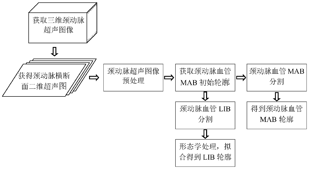
[0061] In the present invention, the carotid artery wall segmentation method based on deep learning in three-dimensional ultrasound images, such as figure 1 shown, including the following steps:

[0062] (1) Obtain three-dimensional carotid artery ultrasound images. The actual three-dimensional carotid ultrasound images of the present invention come from clinical practice. Three-dimensional ultrasound acquisitions were performed on the left and right carotid arteries of 38 patients with carotid artery stenosis exceeding 60%, and a total of 144 three-dimensional carotid artery ultrasound images were obtained.

[0063](2) Cutting the three-dimensional ultrasonic body image into several two-dimensional carotid artery cross-sectional ultrasonic images, and extracting a carotid artery cross-sectional two-dimensional ultrasonic image at intervals of three frames (at this time, the ISD is 4 section planes, two in the present invention) The distance between slice images is 0.1cm), an...


PUM


Abstract

The invention discloses a three-dimensional carotid artery ultrasonic image blood vessel wall segmentation method based on deep learning. The method comprises the following steps: (1) obtaining a three-dimensional ultrasonic image; (2) obtaining a two-dimensional ultrasonic image of a carotid artery cross section, and performing manual marking; (3) dynamically and finely adjusting the convolutional neural network model by utilizing the manually marked image block; (4) fitting vascular adventitia-tunica media boundary initial contour; (5) using the dynamically adjusted convolutional neural network model to carry out vascular adventitia-tunica media boundary contour segmentation; (6) obtaining a vascular cavity ROI region; (7)using U-Net network to divide the vascular cavity, and extractingthe vascular cavity-tunica media boundary contour through morphological processing. According to the method, the contours of vascular adventitia-tunica media boundary MAB and vascular cavity-tunica media boundary LIB can be accurately segmented out; the workload of doctors is greatly reduced, and the vascular wall volume (VWV), the vascular wall thickness (VWT) and the vascular wall thickness change (VWT-Change) can be calculated based on the segmentation result of the method.

Description

technical field [0001] The invention belongs to the intersecting field of computer technology and medical images, and specifically relates to a deep learning-based method for segmenting blood vessel walls in three-dimensional carotid artery ultrasound images. Background technique [0002] In previous studies on ultrasound images of vascular plaques, most of them used two-dimensional B-ultrasound images. Intima-media thickness (IMT) is the most widely used index in clinical evaluation of plaque, which is calculated by calculating the media-adventitia boundary (MAB) and lumen-intima boundary (Lumen- intima Boundary, LIB) to get the distance between. In recent years, three-dimensional ultrasound has provided a more efficient, repeatable, and reliable means of detection and analysis of vascular plaques, which can more reliably analyze the composition, structure, and morphology of plaques while monitoring the effects of drug treatment. Effects of atherosclerosis. Taking the ca...

Claims


Application Information

Patent Timeline
IPC IPC(8): G06T7/13G06T7/11G06T7/155
CPCG06T2207/10136G06T2207/20081G06T2207/20084G06T2207/30101G06T7/11G06T7/13G06T7/155
Inventor 丁明跃周然夏玉娇岳征
Owner HUAZHONG UNIV OF SCI & TECH
Why Eureka
Try Eureka