Focus image recognition method and focus image recognition system based on deep learning model

A technology of image recognition and deep learning, applied in the field of lesion image recognition system, can solve the problem of not reducing the work intensity of doctors, and achieve the effect of avoiding treatment opportunities and reducing work intensity

Active Publication Date: 2019-09-13
HUNAN VATHIN MEDICAL INSTR CO LTD
View PDF11 Cites 26 Cited by
  • Summary
  • Abstract
  • Description
  • Claims
  • Application Information

AI Technical Summary

Problems solved by technology

[0003] In order to solve the problem that the existing medical equipment cannot reduce the work intensity of doctors and detect the disease quickly and early, the purpose of the present invention is to provide a lesion image recognition method and a lesion image recognition system based on a deep learning model

Method used


Image

  • Focus image recognition method and focus image recognition system based on deep learning model
  • Focus image recognition method and focus image recognition system based on deep learning model

Examples


Embodiment 1

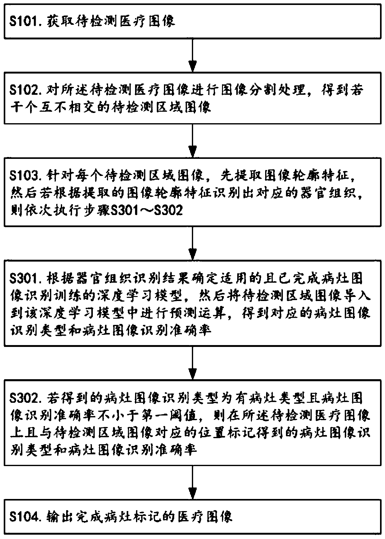
[0045] Such as figure 1 As shown, the lesion image recognition method based on the deep learning model provided in this embodiment includes the following steps S101-S104.

[0046] S101. Acquire a medical image to be detected.

[0047] In the step S101, the medical image to be detected can be specifically derived from an output interface (such as a USB interface or an HDMI interface) of an existing medical photography device. Specifically, the medical images to be detected may be, but not limited to, angiography images, cardiovascular angiography images, computerized tomography images, mammography images, positron emission tomography images, nuclear magnetic resonance imaging images, and medical ultrasound examination images.

[0048] S102. Perform image segmentation processing on the medical image to be detected to obtain several non-intersecting images of regions to be detected.

[0049] In the step S102, since images of multiple organs and tissues are generally included in...

Embodiment 2

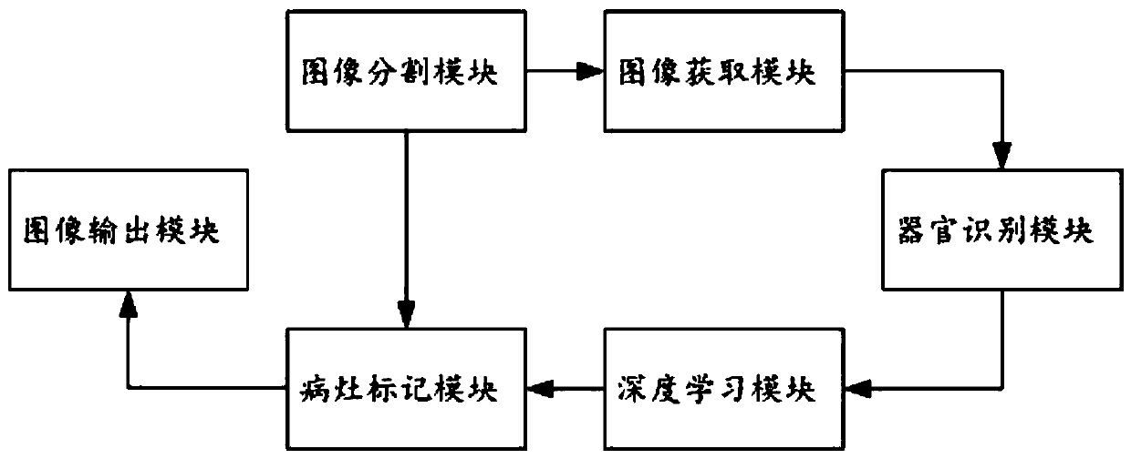
[0063] Such as figure 2As shown, compared with Embodiment 1, this embodiment provides a lesion image recognition system based on the same inventive concept and based on a deep learning model, including an image acquisition module, an image segmentation module, an organ recognition module, a deep learning module, and a lesion image recognition system. A marking module and an image output module; the image acquisition module is used to acquire a medical image to be detected; the image segmentation module is communicatively connected to the image acquisition module, and is used to perform image segmentation processing on the acquired medical image to be detected to obtain Several disjoint images of the region to be detected; the organ identification module is connected to the image segmentation module in communication, and is used to extract the image contour features of the image of the region to be detected, and then identify the corresponding organ tissue according to the extr...


PUM


Abstract

The invention relates to the technical field of medical equipment, and discloses a focus image recognition method and a focus image recognition system based on a deep learning model. The invention provides a new method and a new system for automatically identifying conditions of medical images by replacing doctors based on the deep learning model. Regarding acquired to-be-detected medical images,Segmentation of a smallest organ tissue image and image recognition of a corresponding organ tissue, prediction of a deep learning model and marking on a predication result are carried out. The invention can replace doctors find out underlying conditions of medical images and automatically mark lesion tissues on the medical images. Therefore, a reminder is given to doctors for timely diagnosis. Working intensity of the doctors can be reduced. Whether diseases occur or not can be timely diagnosed. The treatment opportunity of conditions is prevented from being delayed, and the discovery of early-stage diseases is particularly facilitated.

Description

technical field [0001] The invention belongs to the technical field of medical equipment, and in particular relates to a lesion image recognition method and a lesion image recognition system based on a deep learning model. Background technique [0002] At present, the medical imaging equipment in the medical device market can see medical images, such as angiography (Angiography), cardiovascular angiography (Cardiac angiography), computerized tomography (CT, Computerized tomography), mammography ( Mammography), positron emission tomography (PET, Positron emission tomography), nuclear magnetic resonance imaging (NMRI, Nuclear magneticresonance imaging) and medical ultrasound examination images (Medical ultrasound), etc. However, for the discovery or identification of lesions, doctors still use their professional knowledge to judge whether there is a lesion with the naked eye. In the early stage of the lesion, the lesions are often small and easily overlooked. At the same time,...

Claims


Application Information

Patent Timeline
Patent Type & Authority Applications(China)
IPC IPC(8): G06K9/34G06K9/46G06K9/62G06T7/11G06T7/136
CPCG06T7/11G06T7/136G06V10/267G06V10/44G06F18/214
Inventor 不公告发明人
Owner HUNAN VATHIN MEDICAL INSTR CO LTD
Why Eureka
Try Eureka