Skull side surface image analysis method based on neural network and random forest, and system

A side image and neural network technology, applied in the field of computer-aided diagnosis, can solve problems such as shortening the diagnosis cycle, and achieve the effects of shortening the diagnosis cycle, improving stability, high marking accuracy and reliability

Active Publication Date: 2019-09-17
SHANGHAI UEG MEDICAL IMAGING EQUIP CO LTD
View PDF6 Cites 7 Cited by
  • Summary
  • Abstract
  • Description
  • Claims
  • Application Information

AI Technical Summary

Problems solved by technology

[0006] The technical problem to be solved by the present invention is to provide a cranial side image analysis system based on neural network and random forest, which solves the problems existing in the existing automati

Method used


Image

  • Skull side surface image analysis method based on neural network and random forest, and system
  • Skull side surface image analysis method based on neural network and random forest, and system
  • Skull side surface image analysis method based on neural network and random forest, and system

Examples


Embodiment Construction

[0066] Below in conjunction with specific embodiment, further explain the present invention, it should be understood that these embodiments are only used to illustrate the present invention and are not intended to limit the scope of the present invention, after having read the present invention, those skilled in the art will understand various equivalent forms of the present invention All modifications fall within the scope defined by the appended claims of the present application.

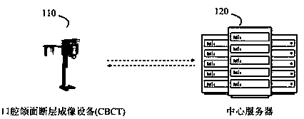
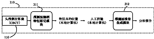
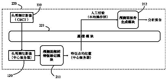
[0067] Such as figure 1As shown, the automatic analysis system of cranial side image based on neural network and random forest disclosed in the embodiment of the present invention uses the imaging device 110 as the data source and the display carrier of the final analysis report, and at the same time uses the central server 120 as an optional auxiliary processing carrier constitute a complete system. The imaging device 110 may be a oral and maxillofacial tomography device (CBCT). In an embodimen...


PUM


Abstract

The invention discloses a skull side surface image analysis method based on a neural network and a random forest, and a system. After imaging equipment (commonly oral and maxillofacial tomography equipment, CBCT in short) finishes patient skull side surface exposure and picture combination, an imaging result is input into a skull side surface dissecting characteristic marking module. Through a series of computer automatic identification and marking processes, marking operation of important dissecting structures on the image is finished, and high-precision positions of characteristic points are output. Afterwards a skull side surface report generating module receives the position of each dissecting characteristic point after artificial verification and adjustment by the skull side surface dissecting characteristic marking module and finishes skull surface medical analysis, wherein a final skull side surface analysis report is used as a subsequent diagnosis basis of the doctor. The method and the system have advantages of settling a problem of performing quick automatic analysis on the skull side surface image, greatly reducing physical labor of a doctor in a skull side surface imaging analysis process, shortening a diagnosis period, improving system predicting stability and preventing abnormal results.

Description

technical field [0001] The invention belongs to the technical field of computer-aided diagnosis, and relates to a method and system for automatic analysis of cranial side X-ray images, in particular to an automatic analysis of cranial side X-ray images (hereinafter referred to as cranial side images) based on random forest technology and neural network technology methods and systems. Background technique [0002] X-ray Cranial Imaging (X-ray Cephalometry) technology is an imaging technology that uses X-rays to project radiation on the patient's head to obtain a side perspective view of the patient's head; its functional feature is to use a small radiation dose, Obtain cross-sectional image data of head bones and tissues to provide basis for clinicians' diagnosis. In the lateral imaging results of the brain, there are some characteristic points indicated by the physiological structure, which can provide auxiliary information for the doctor's diagnosis or become the basis for...

Claims


Application Information

Patent Timeline
IPC IPC(8): G16H50/20G16H30/20G06N3/04
CPCG16H50/20G16H30/20G06N3/045
Inventor 杜鑫陈毅朱露
Owner SHANGHAI UEG MEDICAL IMAGING EQUIP CO LTD
Why Eureka
Try Eureka