Supercharge Your Innovation With Domain-Expert AI Agents!

Clavicle upper arm plexus nerve recognition method and system based on deep learning

A brachial plexus, deep learning technology, applied in the field of neuro-ultrasonic image recognition, can solve problems such as time-consuming, inability to efficiently analyze ultrasonic images, and difficulty in identifying the supraclavicular brachial plexus.

Pending Publication Date: 2019-09-24
王英伟
View PDF0 Cites 0 Cited by
  • Summary
  • Abstract
  • Description
  • Claims
  • Application Information

AI Technical Summary

Problems solved by technology

Due to limited ultrasound resolution, many artifacts, common nerve variations, and the similarity of nerve echoes to those of surrounding connective tissue and fascia, it is difficult to identify the supraclavicular brachial plexus in ultrasound images, and operators need to have extensive experience to do so. Correct identification requires a lot of time and energy to receive relevant training, which is a job that can only be done by senior doctors
At the same time, in the traditional image processing method, the method of processing the image according to some operators is limited by many conditions, resulting in the inability to analyze the ultrasound image accurately and efficiently.

Method used


Image

  • Clavicle upper arm plexus nerve recognition method and system based on deep learning
  • Clavicle upper arm plexus nerve recognition method and system based on deep learning
  • Clavicle upper arm plexus nerve recognition method and system based on deep learning

Examples


Embodiment 1

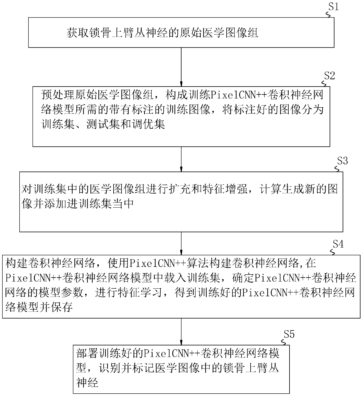
[0061] A recognition method of supraclavicular brachial plexus based on deep learning, such as figure 1 shown, including the following steps:

[0062] Step S1: Obtain a group of original medical image groups of the supraclavicular brachial plexus classified into groups.

[0063] Step S2: Preprocess the original medical image group, crop the original medical image group, and sequentially locate and label the supraclavicular brachial plexus in the original medical image group to form the labeled training image required for training the PixelCNN++ convolutional neural network model , divide the marked images into training set, test set and tuning set.

[0064] Step S3: Expand and enhance the features of the medical image group in the training set, expand and enhance the feature of the medical image group in the training set by adjusting the size, offset, brightness, contrast, grayscale and other parameters of the image, and calculate Generate new images and add them to the trai...

Embodiment 2

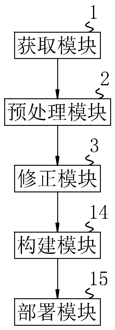
[0083] A recognition system of the supraclavicular brachial plexus based on deep learning, such as figure 2 As shown, the following modules are included:

[0084] Obtaining module 1, used to obtain the original medical image group of the supraclavicular brachial plexus classified into groups;

[0085] Preprocessing module 2, connected with acquisition module 1, is used to preprocess the original medical image group, crop the original medical image group, and sequentially locate and label the supraclavicular brachial plexus in the original medical image group to form the training PixelCNN++ convolutional nerve Annotated training images required by the network model, and the labeled images are divided into training set, test set and tuning set;

[0086] The correction module 3 is connected with the data of the preprocessing module 2, and is used to expand and enhance the features of the medical image group in the training set. By adjusting parameters such as image size, offset...


PUM


Abstract

The invention relates to the technical field of neural ultrasonic image recognition, and discloses a clavicle upper arm plexus nerve recognition method and system based on deep learning, and the method comprises the steps: preprocessing an original medical image of clavicle upper arm plexus nerves, and forming a training image required for training a Pixel CNN + + convolutional neural network model; constructing a convolutional neural network, constructing a convolutional neural network by using a Pixel CNN + + algorithm, loading a training set into the Pixel CNN + + convolutional neural network model, determining model parameters of the Pixel CNN + + convolutional neural network, and deploying a trained Pixel CNN + + convolutional neural network model. The accuracy of recognition can be improved, the recognition speed of subsequent similar images can be increased, and recognition is more accurate and efficient.

Description

technical field [0001] The present invention relates to the technical field of nerve ultrasonic image recognition, and more specifically, it relates to a deep learning-based recognition method and system for the supraclavicular brachial plexus. Background technique [0002] With the rapid development of computer science and technology, people began to use the powerful computing power of computers to realize human recognition skills, especially in medical image processing and analysis and automatic recognition, which played an extremely important role in medical diagnosis. [0003] Ultrasound imaging can display images of the internal tissue structure of the human body in real time and non-invasively. It is very suitable for guiding various anesthesia puncture catheters and targeted drug injection techniques, and has been widely used in clinical anesthesia. When analyzing medical images, the clinician manually analyzes and aims at the target structure in the ultrasound image,...

Claims


Application Information

Patent Timeline
Patent Type & Authority Applications(China)
IPC IPC(8): G06K9/62G06N3/04G16H50/20
CPCG16H50/20G06V2201/03G06N3/048G06N3/045G06F18/214
Inventor 王英伟杨笑宇李根娣郭婷沈雅芳关庆来邹颖华王婷婷
Owner 王英伟
Features
  • R&D
  • Intellectual Property
  • Life Sciences
  • Materials
  • Tech Scout
  • Unparalleled Data Quality
  • Higher Quality Content
  • 60% Fewer Hallucinations
Learn More