Method for intelligently coloring microstructure photo shot by electron microscope and CNN coloring learner

An electron microscope and microstructure technology, applied in instruments, image data processing, computing, etc., can solve the problems of a large number of user parameter input, spend a lot of time and energy, consume knowledge and time, and achieve less training and testing, and faster coloring efficiency. , train and test fast results

Pending Publication Date: 2019-09-27
NINGBO UNIV
View PDF3 Cites 6 Cited by
  • Summary
  • Abstract
  • Description
  • Claims
  • Application Information

AI Technical Summary

Problems solved by technology

At present, the most commonly used method is to use software such as Photoshop, Corel Draw, Fiji, ImageJ, etc. and draw manually. The learning of these software will consume a certain amount of knowledge and time, even if some of these methods provide automatic colorization tools, but they still require a lot of user parameter input
Moreover, these artificial drawing methods need to spend a lot of time and energy for each picture, and if they are handed over to commercial companies, they will need to pay expensive fees
More importantly, such artificial drawing has a great influence on human subjectivity, and the choice of color depends entirely on the painter's preference, which cannot truly reflect the general and conventional colors of the structure.

Method used


Image

  • Method for intelligently coloring microstructure photo shot by electron microscope and CNN coloring learner
  • Method for intelligently coloring microstructure photo shot by electron microscope and CNN coloring learner
  • Method for intelligently coloring microstructure photo shot by electron microscope and CNN coloring learner

Examples


Embodiment 1

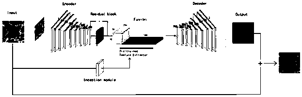
[0043] join figure 1 with 3 , this embodiment provides a method for intelligently coloring end-to-end microstructure photos taken by an electron microscope, including the following steps:

[0044] Step ①: First create a data set, which is a collection of about 1,000 color SEM images. The pictures come from daily life, and the images have different shapes. The shader learns the above data set, and the learning method is based on Google's machine learning Technology, you can refer to the existing literature GoogleResearch.TensorFlow:Large-scale machine learning on heterogeneous systems.Google Res.(2015).doi:10.1207 / s15326985ep4001; Lecun,Y.,Bengio,Y.&Hinton,G.Deep learning.Nature( 2015).doi:10.1038 / nature14539;

[0045] Step ②: Convert the gray-scale photo of the chromosome to be colored taken by the electron microscope into the LAB color space as the input image, L is the brightness, A represents the range from red to green, B represents the range from yellow to blue, and the...

Embodiment 2

[0048] Such as figure 2 with 4 As shown, this embodiment demonstrates a method for intelligently coloring pixel-to-pixel microstructure photos taken by an electron microscope. The method is based on the CNN multi-convolution layer framework and has multiple channels. The three grayscale photos in this embodiment are electron microscope scanning photos of viruses, red blood cells, and neurons from left to right.

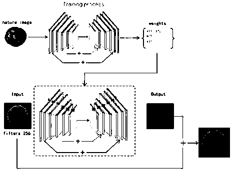
[0049] Manually select a natural color photo in real life as a reference picture for color style transfer, decode the picture and convert its format from RBG to LAB, and obtain the L gray channel Y L and AB color channel Y AB , so as to facilitate the later input to the encoder under the CNN framework of the convolutional neural network. Encoder pair input Y L The color image for coloring output prediction is the predicted channel Y AB ’, the standard Y AB and Y AB ’ The mean square error (MSE) loss generated is passed back to train the convolutional neural ne...

Embodiment 3

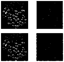
[0053] see Figure 5 , many SEM grayscale images have more repeated similar units. This embodiment uses the grayscale of uniform dense objects in the same reference color image for two different SEM grayscale images of polystyrene nanospheres. The image undergoes end-to-end color style transfer and coloring, and the final output results are in line with the laws of human vision.


PUM


Abstract

The invention discloses a method for intelligently coloring a microstructure photo shot by an electron microscope and a CNN coloring learner, and the method comprises the following steps of firstly, manufacturing a data set which is a set of a plurality of color SEM images, and learning the data set by a shader; converting a to-be-colored microscopic grayscale photo shot by the electron microscope as an input image into an LAB color space, and intercepting a grayscale image with a pixel size of H*W from the microscopic grayscale photo as an input image XL of a CNN; coloring the shader. According to the present invention, the coloring process of the present invention needs to additionally provide a color image in real life similar to the texture structure of a gray image with a colored target as a reference image, and the end-to-end black box type training is conducted on the convolutional neural network, so that the manual participation is not needed.

Description

【Technical field】 [0001] The invention relates to a method for intelligently coloring microstructure photos taken by an electron microscope and a CNN coloring learner, belonging to the field of image coloring of electron microscope pictures. 【Background technique】 [0002] After the invention of the scanning electron microscope (SEM) in 1937, nanoscience, nanotechnology experiments and their observation became relatively easy. It gives us some great images at the nanometer or micrometer level. SEM imaging has made great contributions to different researchers and is widely used in nanotechnology and nanoscience. Nanotechnology is nanoscale (10 -9 m) The cutting-edge science of manipulating objects to create new and unique materials and products. The SEM images of nanotechnology fields such as graphene, protein, two-dimensional materials and any microelectronic experiments are presented to everyone in the form of black and white photos. It has promoted the rapid developmen...

Claims


Application Information

Patent Timeline
Patent Type & Authority Applications(China)
IPC IPC(8): G06T3/00
CPCG06T3/0012G06T2207/10061G06T2207/20084G06T2207/20081G06T2207/10024G06T2207/20221
Inventor 林冬冬伊斯莱尔·戈伊托姆·比日哈尼王钦谢正
Owner NINGBO UNIV
Why Eureka
Try Eureka