Method for constructing virtual physiological tissues of sinus node, storage medium and computing device

A construction method and tissue technology, applied in the field of translational medicine, can solve the problems that the atrial model cannot reproduce early afterdepolarization and late afterdepolarization, lacks experimental data, and does not have the physiological functions of human cardiomyocytes.

Active Publication Date: 2019-10-08
JINAN UNIVERSITY
View PDF7 Cites 0 Cited by
  • Summary
  • Abstract
  • Description
  • Claims
  • Application Information

AI Technical Summary

Problems solved by technology

The following problems exist in the study of the human sinus node: 1) The difference between the current microscopic research and the macroscopic research makes people lack a systematic understanding of the function of the sinus node; 2) Considering ethical issues, there is a lack of human sinus node cells, tissues and structures 3) There are great differences between the sinus nodes of animals and humans, and the availability and reliability of animal experiments on human sinus nodes are limited. dispute
[0005] The prior art document Computational assessment of the functional role of sinoatrial node exit pathways in the human heart (Kharche S R, Vigmond E, Efimov I R, et al. [J]. PloS one, 2017, 12 (9): e0183727.) To the construction method of sinoatrial node tissue, but this method has the following defects: (1) The whole tissue structure includes central sinoatrial node tissue, peripheral sinoatrial node tissue and atrium, and does not contain non-exciting tissues (such as border ridge or fat, etc.) , therefore, the representation of the tissue structure is insufficient; (2) All cells in the tissue adopt the Fenton-Karma calculation model, which is not constructed based on human experimental data and does not have the physiological functions of human cardiomyocytes: the virtual physiological tissue does not contain Calcium clock, atrial model cannot reproduce early afterdepolarization and late afterdepolarization in biological experiments

Method used


Image

  • Method for constructing virtual physiological tissues of sinus node, storage medium and computing device
  • Method for constructing virtual physiological tissues of sinus node, storage medium and computing device
  • Method for constructing virtual physiological tissues of sinus node, storage medium and computing device

Examples


Embodiment 1

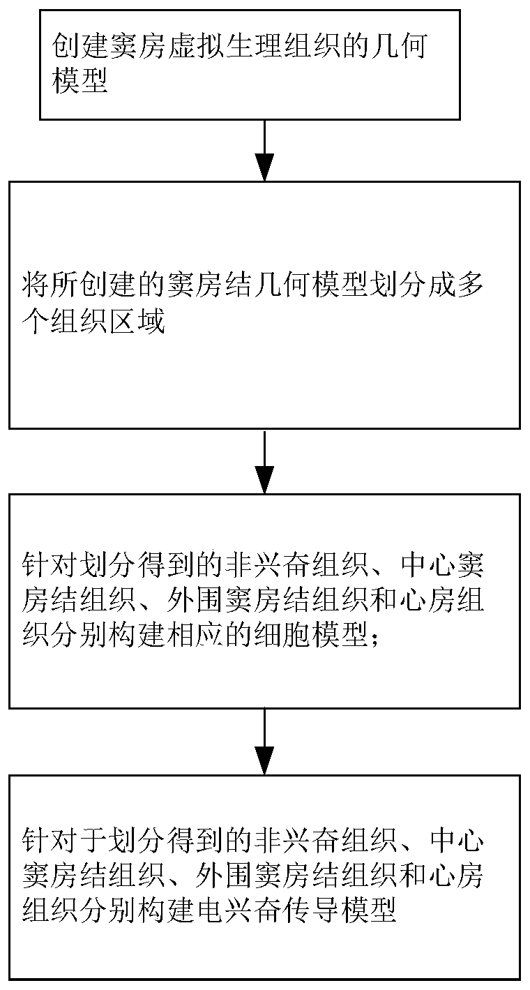
[0073] The invention discloses a method for constructing a virtual physiological tissue of the sinus node. The constructing method is realized in a computer, such as figure 1 As shown, including the following steps:

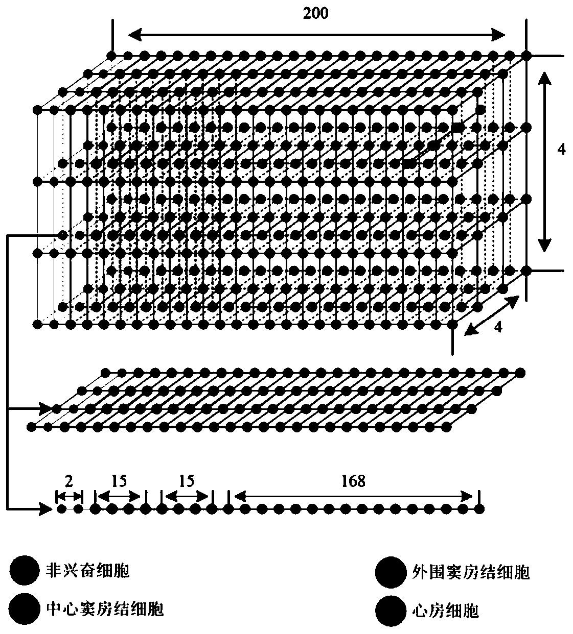
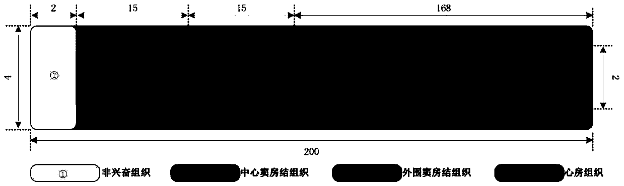
[0074] S1. Create a geometric model of virtual physiological tissue; in this embodiment, such as Figure 2a with 2b As shown, the geometric model of the virtual physiological tissue is a rectangular parallelepiped, and the number of nodes in the length, width and height are 200, 4 and 4 respectively; each two adjacent nodes in the length, width and height are separated by a certain distance , Called the space step; each node represents a cell.

[0075] S2. Divide the created sinus node virtual physiological tissue geometric model into multiple areas, including non-excited tissue area, central sinus node tissue area, peripheral sinus node tissue area and atrial tissue area;

[0076] Such as Figure 2a with 2b As shown, in this embodiment, in the sinus node virtual physi...

Embodiment 2

[0130] This embodiment discloses a storage medium that stores a program, and when the program is executed by a processor, the method for constructing a virtual physiological tissue of the sinus node described in Embodiment 1 is implemented, which is specifically as follows:

[0131] Create a geometric model of the sinoatrial virtual physiological tissue;

[0132] Divide the created sinoatrial node geometric model into multiple areas, including non-excited tissue area, central sinus node tissue area, peripheral sinus node tissue area, and atrial tissue area;

[0133] For the divided non-excited tissue, central sinoatrial node tissue, peripheral sinoatrial node tissue and atrial tissue, corresponding cell models were constructed:

[0134]

[0135] Among them, for non-excited tissues, the first cell model was constructed using a sinoatrial node cell model that does not contain L-type calcium ion current: Where C m1 Indicates the cell membrane capacitance of non-excited tissue; V 1 Repres...

Embodiment 3

[0155] The present invention discloses a computing device, which includes a processor and a memory for storing an executable program of the processor. When the processor executes the program stored in the memory, the method for constructing the virtual physiological tissue of the sinus node in embodiment 1 is implemented. as follows:

[0156] Create a geometric model of the sinoatrial virtual physiological tissue;

[0157] Divide the created sinoatrial node geometric model into multiple areas, including non-excited tissue area, central sinus node tissue area, peripheral sinus node tissue area, and atrial tissue area;

[0158] For the divided non-excited tissue, central sinoatrial node tissue, peripheral sinoatrial node tissue and atrial tissue, corresponding cell models were constructed:

[0159]

[0160] Among them, for non-excited tissues, the first cell model was constructed using a sinoatrial node cell model that does not contain L-type calcium ion current: Where C m1 Indicates t...


PUM


Abstract

The invention discloses a method for constructing virtual physiological tissues of a sinus node, a storage medium and a computing device. The method comprises the following steps: creating a geometricmodel for the virtual physiological tissues of a sinus; dividing the geometric model into a plurality of areas including a non-excited tissue area, a central sinus node tissue area, a peripheral sinus node tissue area and an atrium tissue area; respectively constructing corresponding cell models for the non-excited tissues, the central sinus node tissues, the peripheral sinus node tissues and theatrium tissues; and respectively constructing electrical excitation conduction models for the non-excited tissues, the central sinus node tissues, the peripheral sinus node tissues and the atrium tissues obtained after the division. The virtual physiological tissues constructed by the method of the invention construct a bridge for changing from microscopic molecules to macroscopic organ, recurring sinus node pace-making and electrical conduction processes are in line with the electrophysiology of the human sinus node, the time and money cost of animal experiments is reduced, and the pace-making mechanism of the sinus node can be fast, well and safely studied.

Description

Technical field [0001] The invention relates to the technical field of translational medicine, in particular to a method for constructing a virtual physiological tissue of the sinus node, a storage medium and a computing device. Background technique [0002] The sinus node is the pacing point of the human heart. It is located in the upper right atrium, near the upper end of the sulcus where the right atrium and the superior vena cava junction. The size of the adult sinoatrial node is 15mm×5mm×2mm. The sinoatrial node is flat, surrounded by non-excitable tissue and non-autonomous atrial tissue, and inside is sinus node cells, which are composed of central cells (starting from Stroke cells) and peripheral cells (transition cells). The central cell is small and is the place where cardiac pacing occurs; the peripheral cell is the connecting cell between the central cell and the atrial cell. Its structure and morphology are between the central cell and the atrial cell and are respons...

Claims


Application Information

Patent Timeline
Patent Type & Authority Applications(China)
IPC IPC(8): G16H50/50
CPCG16H50/50
Inventor 白杰云陆尧胜杨仁桓
Owner JINAN UNIVERSITY
Why Eureka
Try Eureka