Preparation method of individualized 3D printed high tibial osteotomy guide plate

A 3D printing and osteotomy technology, used in bone drill guidance, medical science, surgery, etc., can solve the problems of difficulty in ensuring the accuracy of the osteotomy position, increasing the risk of postoperative infection for patients, and easily endangering the health of surgeons. The effect of shortening surgical anesthesia time, shortening operation time, and ensuring safety

Active Publication Date: 2019-11-01
XIAN HONGHUI HOSPITAL
View PDF17 Cites 11 Cited by
  • Summary
  • Abstract
  • Description
  • Claims
  • Application Information

AI Technical Summary

Problems solved by technology

The entire osteotomy positioning process is cumbersome, prolongs the entire operation time, and increases the risk of postoperative infection for patients
Moreover, the accuracy of positioning is greatly affected by the human factors of the surgeon, and it is difficult to ensure the accuracy of the osteotomy position determination, and there are unfavorable factors of radiation and high radiation during the fluoroscopy process, which is likely to endanger the health of the surgeon.
During the entire osteotomy process, positioning is based entirely on the experience of the surgeon and the naked eye, and precise control cannot be achieved, and the shape and length of the proximal tibia are different for each patient. How to perform personalized and precise osteotomy is a further problem to be solved.

Method used


Image

  • Preparation method of individualized 3D printed high tibial osteotomy guide plate
  • Preparation method of individualized 3D printed high tibial osteotomy guide plate

Examples


Embodiment Construction

[0027] The technical solutions of the present invention will be further described in detail below in conjunction with the embodiments.

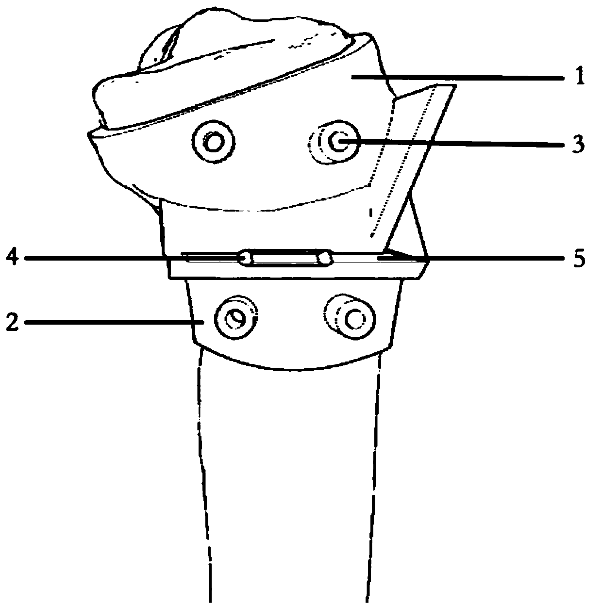
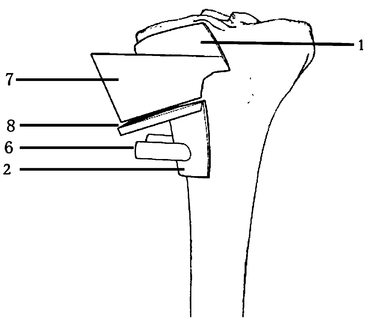
[0028] Such as figure 1 As shown, this embodiment provides an osteotomy guide plate for high tibial osteotomy. This osteotomy guide is designed in one-piece 3D printing. figure 1 The middle and upper plate body 1 and the lower plate body 2 are integrally connected; the positioning hole 3 at the proximal end of the tibia is the position where the osteotomy guide is fixed to the patient’s tibia with a 2mm diameter Kirschner wire, and the four fixing holes are short post holes It is quadrilateral and distributed parallel to the upper and lower plates; the Kirschner wire hole 4 of the guide is the initial stabilization device of the osteotomy guide plate for high tibial osteotomy, and it is a supplementary device for the positioning hole 3 at the proximal end of the tibia. Drive into the guide hole of the calibration shaft, and after the osteoto...


PUM


Abstract

The invention discloses a preparation method of an individualized 3D printed high tibial osteotomy guide plate, for an osteotomy guide plate of a 3D printed high tibial osteotomy. Before a surgery, according to the CT data of the knee joint of a patient, through computer simulation modeling and a 3D reconstruction procedure, the deformity shape of the tibia shape of a patient is printed in proportion being 1 to 1, an osteotomy position is designed before the surgery through a 3D printing technique, and finally an osteotomy guider which is completely fitted to the near end of the tibia of the patient is designed through a model material; and in the surgery process, the osteotomy guider is directly fitted to the near end of the tibia in the surgery process, according to the preoperative osteotomy scheme, osteotomy surgery operations are immediately started, under regulation for guaranteeing accuracy, the surgery operation steps are simplified, the anaesthesia time of the surgery is shortened, the safety of the surgery of the patient is guaranteed, and the preparation method has great clinical promotion effects. A steel plate can be fixed according to tibia near ends provided by different factories, the position and the direction of the osteotomy of the tibia can be in individualized design, and the preparation method has definite diversified flexibility.

Description

technical field [0001] The invention belongs to the technical field of orthopedic medical devices, and relates to a method for preparing a personalized 3D printed high tibial osteotomy guide plate, in particular, to a preparation and preparation of a personalized 3D printed osteotomy guide plate for high tibial osteotomy. How to use it. Background technique [0002] Degenerative osteoarthropathy of the knee joint is often accompanied by varus deformity of the knee joint, and produces changes in the load-bearing biological stress in the joint, which leads to an imbalance in the force line of the lower limbs, which in turn accelerates the wear of the knee articular cartilage and the development of bone joints. At present, high medial tibial osteotomy is the most common surgical method for the treatment of early and mid-stage knee osteoarthritis caused by knee varus. The principle of the treatment is to transfer the pressure of the worn medial compartment of the knee joint to ...

Claims


Application Information

Patent Timeline
Patent Type & Authority Applications(China)
IPC IPC(8): A61B17/17B33Y30/00B33Y50/00
CPCA61B17/1732A61B17/1739B33Y30/00B33Y50/00
Inventor 张宪任博王富民
Owner XIAN HONGHUI HOSPITAL
Why Eureka
Try Eureka