Correction information acquisition method for performing attenuation correction on PET images of breath or heart

A technology of attenuation correction and correction information, which is applied in the field of PET systems, can solve the problems of attenuation image truncation, increased workload of doctors, and low signal-to-noise ratio of gated images, so as to improve image signal-to-noise ratio, solve attenuation artifacts, and improve The effect of image quality

Active Publication Date: 2021-11-16
JIANGSU SINOGRAM MEDICAL TECH CO LTD
View PDF6 Cites 0 Cited by
  • Summary
  • Abstract
  • Description
  • Claims
  • Application Information

AI Technical Summary

Problems solved by technology

[0006] 1. After PET data acquisition, each phase is reconstructed separately, so the data available in the reconstruction process is much less than that of static scanning, which leads to a very low signal-to-noise ratio of the final gated image
In this way, even if the blurring effect of object motion on the lesion is removed, the added noise will greatly reduce the detectability of small lesions
[0007] 2. Since the other modality images correspond to a certain phase position, only one phase in the PET multi-phase image obtained by the gating method can match the anatomical structure image, which will still cause the PET image to be matched with other modality images. Quasi-fusion produces deviations (especially near the diaphragm with the largest movement range), and using instantaneous multi-modal images to perform attenuation correction on the average PET image will inevitably cause errors, which will produce local artifacts on the PET image and affect quantitative analysis. , may affect the accurate diagnosis and treatment plan formulation of thoracic and abdominal tumors
When scanning heavy patients, imaging with other modalities will likely not provide a large enough scan field, resulting in truncated attenuation images
This incomplete attenuation information can also produce attenuation artifacts in PET reconstructions
[0009] 4. When PET is combined with other phantoms, sometimes satisfactory attenuation-corrected images cannot be obtained, such as PET / MR imaging
On the one hand, this increases the workload of doctors. On the other hand, only one frame of images appears in the diagnostic report, and other reconstruction results are not used. This has a lot of information redundancy, wasting data and computing time

Method used


Image

  • Correction information acquisition method for performing attenuation correction on PET images of breath or heart
  • Correction information acquisition method for performing attenuation correction on PET images of breath or heart
  • Correction information acquisition method for performing attenuation correction on PET images of breath or heart

Examples


Embodiment 1

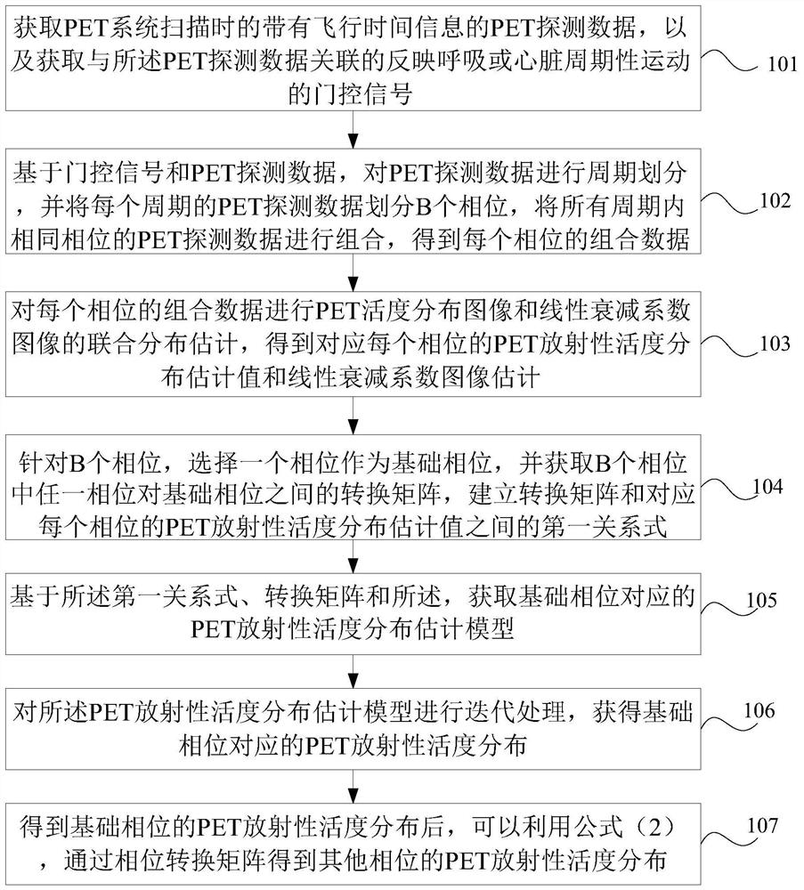
[0082] The present application proposes a correction information acquisition method for attenuation correction of PET images of respiration or heart. like figure 1 The specific steps of this method are as follows:

[0083] 101. Acquire PET detection data (hereinafter referred to as PET data) with time-of-flight information when the PET system scans, and acquire a gated signal that is associated with the PET detection data and reflects respiration or cardiac periodic motion.

[0084] In the specific implementation process, the gate control signal can be extracted through an external gate control device. Alternatively, in a possible implementation, the above-mentioned PET data itself is used to extract the gating signal to reflect the periodic motion of respiration or the heart.

[0085] 102. Based on the gating signal and the PET detection data, divide the PET detection data into cycles, divide the PET detection data of each cycle into B phases, and perform the PET detection ...

Embodiment 2

[0122] An embodiment of the present invention provides a correction information acquisition method for performing attenuation correction on a PET activity distribution image. The method includes the following steps:

[0123] S0. Acquire PET detection data and other modal images with time-of-flight information when the PET system scans.

[0124] For example, other modality images may include: CT images or MR images.

[0125] S1. Based on the Poisson distribution of the PET detection data, modeling processing is performed on the PET detection data to obtain the log-likelihood function L(x, μ, y) of formula (p1);

[0126]Formula (p1)

[0127] Among them, y=[y 1t ,y 2t ,…,y NT ] T represents the detection data, N represents the size of the sinogram of the detection data, T represents the dimension of the TOF of the flight time; x=[x 1 ,x 2 ,…,x J ] T Represents the unknown PET radioactivity distribution, J represents the size of the discrete space of the PET image; μ=[μ...

Embodiment 3

[0168]The present invention also provides a PET image reconstruction method, which includes:

[0169] M01. Obtain the output value of the PET radioactivity distribution x corresponding to the basic phase by using any of the methods described in Embodiment 1 above;

[0170] M02. According to the output value of the PET radioactivity distribution x corresponding to the basic phase, apply the output value in the reconstruction of the PET activity distribution image scanned by the PET system to obtain a PET image of the basic phase.

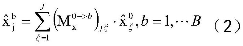
[0171] In another embodiment, any method described in Embodiment 1 above can also be used to obtain the output value of the PET activity distribution x corresponding to any phase in the cycle, and then apply it to the PET activity distribution scanned by the PET system In image reconstruction, a PET image of each phase can be obtained.

[0172] In addition, the PET images of each phase can also be combined and processed to obtain a period of reconst...


PUM


Abstract

The invention discloses a correction information acquisition method for performing attenuation correction on PET images of breathing or heart. The method includes: acquiring PET detection data with time-of-flight information and gating signals reflecting periodic movement of breathing or heart; The PET detection data of a cycle is divided into B phases, the PET detection data of the same phase in all cycles are combined, the sum of each phase is obtained, and the transformation matrix between any phase in the B phases and the basic phase is obtained, and the transformation is established. The first relationship between the matrix and the estimated value of the PET activity distribution corresponding to each phase; obtain the PET activity distribution estimation model corresponding to the basic phase; iteratively process the PET activity distribution estimation model to obtain The PET activity distribution corresponding to the basal phase. The foregoing can solve the problem of attenuation artifacts in PET image reconstruction in the prior art, and can ensure accurate quantification of attenuation correction.

Description

technical field [0001] The invention relates to the field of medical imaging, in particular to a correction information acquisition method and a PET system for performing attenuation correction on PET images of respiration or heart. Background technique [0002] Positron Emission Tomography (PET) is a high-end nuclear medicine imaging diagnostic equipment. Use of radionuclides (such as 18 F. 11 C, etc.) Label metabolites and inject nuclides into the human body, and then perform functional metabolic imaging on patients through PET system to reflect the metabolic activities of life. PET scans are usually matched with other modal scans (such as CT, MRI, etc.) to obtain the imaging of the patient's anatomical structure. The tissue density distribution of the patient is obtained, the attenuation property (attenuation coefficient) of the tissue to the radiation is calculated, and then applied to the image reconstruction, the attenuation correction is performed on the PET data, ...

Claims


Application Information

Patent Timeline
Patent Type & Authority Patents(China)
IPC IPC(8): G06T5/00G06T7/00G06T11/00
CPCG06T5/006G06T7/0012G06T11/005G06T2207/10104G06T2207/30048
Inventor 李楠
Owner JIANGSU SINOGRAM MEDICAL TECH CO LTD
Why Eureka
Try Eureka