A method for automatic choroidal segmentation of OCT images based on gcs-net

An automatic segmentation and choroid technology, applied in image analysis, image enhancement, image data processing, etc., can solve problems such as difficulties, single local information, complex detection, etc., and achieve the effect of enhancing consistency

Active Publication Date: 2021-10-19
SUZHOU UNIV
View PDF3 Cites 0 Cited by
  • Summary
  • Abstract
  • Description
  • Claims
  • Application Information

AI Technical Summary

Problems solved by technology

[0004] At present, both traditional algorithms and deep learning can segment the choroid in OCT images, but both have certain limitations and deficiencies: (1) The traditional algorithm is complex and the detection accuracy of the subchoroidal boundary is not high
(2) Many traditional algorithms are only applicable to normal retina, or only to images centered on the macula. For pathological myopia or retina with optic nerve head (ONH), the detection becomes more complicated and difficult
(3) Although deep learning can make up for the shortcomings of traditional algorithms, most of the existing networks uniformly process all feature maps of the same layer, resulting in the same receptive field of the network in the same layer, thus obtaining a single local information
(4) With the continuous downsampling of the network and the convolution operation with a step size, the defect that only a single size of information can be obtained at the same layer becomes more and more obvious, resulting in inaccurate segmentation of the choroid

Method used


Image

  • A method for automatic choroidal segmentation of OCT images based on gcs-net
  • A method for automatic choroidal segmentation of OCT images based on gcs-net
  • A method for automatic choroidal segmentation of OCT images based on gcs-net

Examples


Embodiment

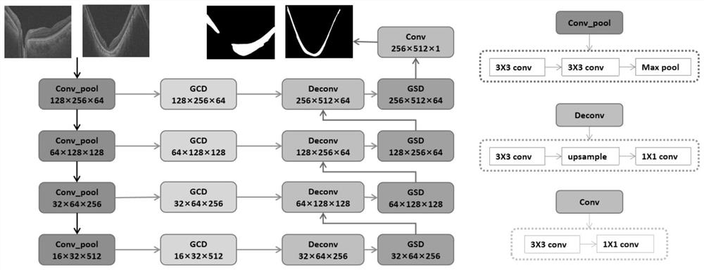
[0045] 1) Data preprocessing

[0046] The experimental data set is composed of large-field three-dimensional OCT images collected by a Topcon DRI-OCT scanner with a central wavelength of 1050 nm, and the scanning range includes the center of the macula and the optic nerve head (ONH) area. The image size is 512(B-scan width)×256(B-scan times)×992(B-scan depth), and the corresponding volume is 12×9×2.6mm 3 . The data set consists of 1650 gold-standard B-scan OCT images marked by professional doctors, of which 1150 are from 115 normal human eyes, 500 are from 50 highly myopic human eyes, and 10 evenly spaced B-scans are extracted from each human eye. Scan OCT images.

[0047] In the process of training and verification, in order to improve the computational efficiency of the model, the original OCT image with a size of 512 (B-scan width) × 992 (B-scan depth) was bilinearly interpolated and down-sampled to 256 (B-scan width) × 512 ( B scan depth), and randomly flip the image le...


PUM


Abstract

The invention discloses a method for automatically segmenting the choroid of OCT images based on GCS-Net. Between each layer across the connection layer, the inter-group channel dilation module (GCD) is used for connection, and the inter-layer connection is made by the inter-group space dilation module (GSD) after each deconvolution layer in the decoding path; the trained GCS ‑Net network model for testing, input the image to be segmented into the built model, and output the corresponding choroidal segmentation map. The above two modules adopt two methods to automatically select multi-scale information between groups, which significantly improves the accuracy of automatic choroid segmentation, and the applicable objects can be extended to pathological myopia or retinal images containing optic nerve head. The present invention is beneficial to improve The accuracy of the quantitative analysis of the choroid is also conducive to the comprehensive acquisition of the morphological information of the choroid in the three-dimensional large field of view data.

Description

technical field [0001] The invention relates to a method for automatically segmenting the choroid of an OCT image based on GCS-Net, and belongs to the technical field of fundus image segmentation. Background technique [0002] The choroid is a layer of complex vascular system between the retinal pigment epithelium (RPE) and the sclera, which has very important physiological functions. The distribution of choroidal thickness and changes in volume in OCT images have become important indicators for the management of retinal diseases. Many diseases are closely related to the morphology of the choroid, such as pathological myopia (PM), glaucoma, age-related macular degeneration (AMD), central serous chorioretinopathy (CSC), myopic maculopathy, choroiditis, etc. Realizing the automatic segmentation of the choroid in OCT images is of great significance for detecting early lesions, observing the course of the disease, and studying pathology. [0003] Swept-frequency optical cohere...

Claims


Application Information

Patent Timeline
Patent Type & Authority Patents(China)
IPC IPC(8): G06T7/00G06T7/11
CPCG06T7/0012G06T2207/20081G06T2207/20084G06T2207/30041G06T7/11
Inventor 石霏陈新建成雪娜朱伟芳
Owner SUZHOU UNIV
Why Eureka
Try Eureka