Segmentation method of cardiac structure in MRI images based on multi-channel convolutional neural network

A technology of convolutional neural network and heart structure, applied in the field of medical image processing, can solve the problems of high computational cost, thick scanning layer, large spacing, etc., to achieve the effect of improving precision and accuracy, improving segmentation performance, and high computational cost

Active Publication Date: 2022-04-19
SICHUAN UNIV
View PDF8 Cites 0 Cited by
  • Summary
  • Abstract
  • Description
  • Claims
  • Application Information

AI Technical Summary

Problems solved by technology

The method based on deep learning can obtain more accurate automatic segmentation results. However, the existing heart segmentation methods based on deep learning mostly use 2D segmentation methods without considering the inter-layer context information. The inter-layer context information is important for accurate segmentation and improved segmentation. performance is valuable
Ignoring inter-layer contextual information is not in line with the actual workflow of clinicians
At the same time, due to the thick and large spacing of the scanning layers of the cardiac cine MRI image, directly using the inter-layer context information through the 3D segmentation method is not only computationally expensive but may not bring about performance improvement.

Method used


Image

  • Segmentation method of cardiac structure in MRI images based on multi-channel convolutional neural network
  • Segmentation method of cardiac structure in MRI images based on multi-channel convolutional neural network
  • Segmentation method of cardiac structure in MRI images based on multi-channel convolutional neural network

Examples


Embodiment Construction

[0030] In order to make the object, technical solution and advantages of the present invention clearer, the present invention will be further described in detail below in combination with specific embodiments and with reference to the accompanying drawings. It should be understood that these descriptions are exemplary only, and are not intended to limit the scope of the present invention. Also, in the following description, descriptions of well-known structures and techniques are omitted to avoid unnecessarily obscuring the concept of the present invention.

[0031] The cardiac cine MRI image in the present invention refers to an image obtained by cardiac magnetic resonance cine imaging technology, which is a kind of cardiac MRI image.

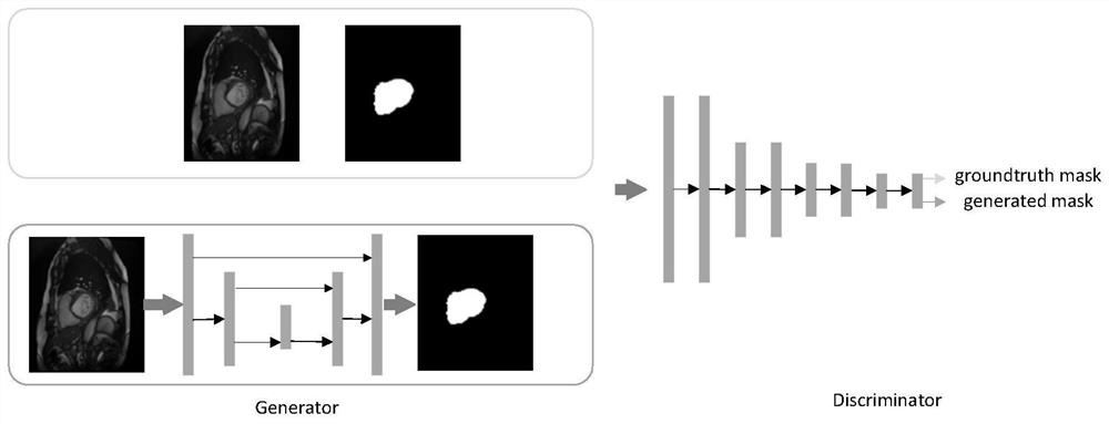
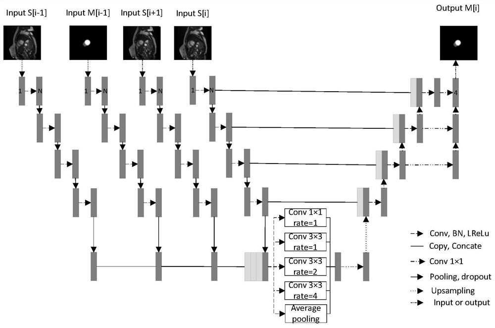
[0032] The ASPP module in the present invention refers to a pyramid pooling module with dilated convolution.

[0033] Cardiac magnetic resonance cine imaging technology is a commonly used cardiac magnetic resonance imaging technology, which u...


PUM


Abstract

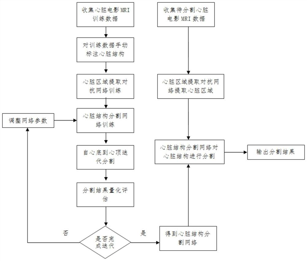
The present invention relates to a method for segmenting cardiac structures in MRI images based on a multi-channel convolutional neural network, which includes collecting heart movie MRI training data of normal people and heart patients, and manually marking the cardiac structures in the training data by experienced doctors as Heart segmentation labeling results, based on the training data to train the heart region extraction model, so that the heart region extraction model can accurately extract the heart region, and train the heart segmentation network according to the heart region extracted from the training data to segment the heart Structure, using the standard segmentation labeling results as a standard to measure the segmentation performance of the constructed heart segmentation network. The present invention uses a heart region extraction model based on a generative confrontation network to extract the heart, which improves the accuracy of the heart region extraction; at the same time, the context information between adjacent layers is used through a multi-channel convolutional neural network, which improves the segmentation accuracy and accuracy. Spend.

Description

technical field [0001] The invention relates to the field of medical image processing, in particular to a method for segmenting cardiac structures in MRI images based on a multi-channel convolutional neural network. Background technique [0002] According to the statistics of the World Health Organization, cardiovascular disease is the disease with the highest mortality rate in the world, and about 19.7 million people died of cardiovascular disease in 2016. In clinical practice, cardiac function analysis plays an important role in heart disease diagnosis, risk assessment, patient management, and treatment decision-making. This is usually done with the aid of digital images of the heart to quantify global or regional cardiac function by assessing a range of clinical parameters such as ventricular volume, ejection fraction, stroke volume, myocardial mass, etc. Due to the good discrimination of soft tissues, the evaluation of left and right ventricular ejection fraction, strok...

Claims


Application Information

Patent Timeline
Patent Type & Authority Patents(China)
IPC IPC(8): G06T7/11G06T7/174G06N3/04G06N3/08
CPCG06T7/11G06T7/174G06N3/08G06T2207/10088G06T2207/20076G06T2207/20221G06T2207/30048G06N3/045
Inventor 马宗庆吴锡
Owner SICHUAN UNIV
Why Eureka
Try Eureka