
Nonenzymatic biosensor for detecting activity of uracil-DNA glycosylase

A biosensor and glycosylase technology, applied in the field of biosensors, can solve the problems of complex operation, high cost, low specificity and sensitivity, etc., and achieve the effect of good repeatability, simple operation and good specificity

Active Publication Date: 2020-01-31
FUZHOU UNIV
View PDF2 Cites 5 Cited by
  • Summary
  • Abstract
  • Description
  • Claims
  • Application Information

AI Technical Summary

Problems solved by technology

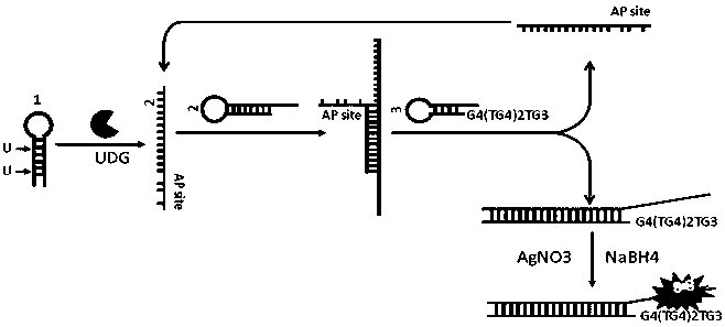
[0003] In order to solve the problems of relatively low specificity and sensitivity, high cost and complicated operation in the method for detecting uracil-DNA glycosylase in the above prior art, the present invention provides a detection method with high specificity and sensitivity, low cost and Fast biosensing based on catalytic hairpin assembly amplification and silver nanocluster fluorescence intensity change detection of uracil-DNA glycosylase

Method used


Image

  • Nonenzymatic biosensor for detecting activity of uracil-DNA glycosylase
  • Nonenzymatic biosensor for detecting activity of uracil-DNA glycosylase
  • Nonenzymatic biosensor for detecting activity of uracil-DNA glycosylase

Examples


Embodiment 1

[0035] Example 1 Preparation of DNA Templated Silver Nanoclusters

[0036] Formulating AgNO 3 The concentration is 100 μM, the volume is 1 mL, and stored in the dark at 4°C. Prepare NaBH 4Concentration 100 μM, volume 1 mL, NaBH 4 It is ready-to-use and prepared with ice water at 0°C.

[0037] The solution after catalytic hairpin self-assembly contains 2 strands (5 μL, 10 μM), so add the prepared AgNO 3 (100μM) solution 3.0μL, after shaking for 30s, place it in a dark place at 4°C for 20min, then take it out and add freshly prepared NaBH 4 (100 μM) solution 3.0 μL, after shaking for 30 s, place at 4 °C in the dark for 3 h.

Embodiment 2

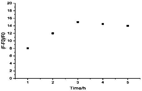
[0038] Example 2 Fluorescence intensity changes with incubation time.

[0039] (1) Mix 2 μL of chain 1 (5 μM), 1 μL of UDG (100 U / mL) and 10 μL of UDG-10×Reaction Buffer, and react at 37°C for 1 hour. The UDG-10×Reaction Buffer: contains 20 mM Tris-HCl (pH=8.2, 25° C.), 10 mM EDTA, and 100 mM NaCl.

[0040] (2) Heat the solution in step (1) at 90°C to inactivate UDG enzyme, then cool it down to 20°C for later use.

[0041] (3) Add 10 μL of 2-strand (5 μM) and 10 μL of 3-strand (5 μM) to the solution in step (2) to carry out catalytic hairpin self-assembly reaction at 20° C. for 2 h.

[0042] (4) Add AgNO to the solution in step (3) 3 (100μM) solution 3.0μL, after shaking for 30s, place it in a dark place at 4°C for 20min, then take it out and add freshly prepared NaBH 4 (100 μM) solution 3.0 μL, shake for 30 seconds, and add 20 mM PBS buffer to make the final reaction volume 100 μL, incubate at 4°C in the dark, and the incubation time is 1h, 2h, 3h, 4h, 5h respectively. Fl...

Embodiment 3

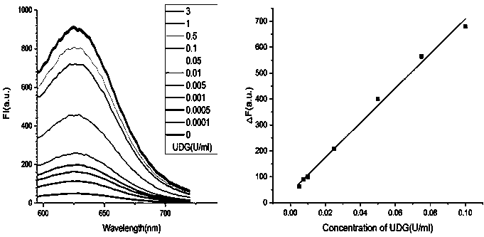
[0044] Example 3 Activity detection of UDG

[0045] (1) Mix 2 μL of chain 1 (5 μM) with different concentrations of UDG (the final concentration of UDG is 0 U / mL, 0.0001 U / mL, 0.0005 U / mL, 0.001 U / mL, 0.005 U / mL, 0.01U / mL, 0.05U / mL, 0.1U / mL, 0.5U / mL, 1U / mL, 3U / mL) and 10μL of UDG-10×Reaction Buffer were mixed and reacted at 37°C for 1h. The final volume of the reaction was 100 μL.

[0046] (2) Heat the solution in step (1) at 90°C to inactivate UDG enzyme, then cool it down to 20°C for later use.

[0047] (3) Add 10 μL of 2-strand (5 μM) and 10 μL of 3-strand (5 μM) to the solution in step (2) to carry out catalytic hairpin self-assembly reaction at 20°C for 2 h.

[0048] (4) Add AgNO to the solution in step (3) 3 (100μM) solution 3.0μL, after shaking for 30s, place it in a dark place at 4°C for 20min, then take it out and add freshly prepared NaBH 4 (100 μM) solution 3.0 μL, shake for 30 seconds, and add 20 mM PBS buffer to make the final reaction volume 100 μL, incubate ...


PUM


Abstract

The invention provides a nonenzymatic biosensor for detecting activity of uracil-DNA glycosylase. The nonenzymatic biosensor comprises a UDG template strand 1, a silver nanocluster DNA template strand2, and a DNA strand 3 rich in cytosine. A fluorescence method is used for detecting the activity of the uracil-DNA glycosylase, the stimulation wave lenagth of fluoroscopic examination is 570nm, thelength of transmitted waves is 630nm, and the detection wave band is 590-720nm. The biosensor provided by the invention is good in specificity, high in sensitivity, mild in reaction condition and highin reaction speed. Because the reaction process is free from addition of external enzymes, and through silver nanocluster fluoroscopic examination, the biosensor is simple to operate, low in reactionrequirements, short in detection cycle, stable in properties, and good in repeatability, and is suitable for detection of UDG in the field of medical health.

Description

technical field [0001] The invention belongs to the technical field of biosensors, and relates to an enzyme-free biosensor for catalyzing hairpin self-assembly amplification and silver nano-cluster fluorescence intensity change detection for uracil-DNA glycosylase. Background technique [0002] In living organisms, the integrity of genetic information is constantly threatened by reactive free radical species, toxins, and radiation, which lead to the conversion of cytosine bases into uracil bases in the genome, causing a high degree of DNA base mutagenic damage. Uracil-DNA glycosylase (UDG) is one of the most important DNA repair enzymes (BER), which plays an important repair role. UDG specifically removes damaged uracil bases in double-stranded and single-stranded DNA by hydrolyzing the glycosidic bonds between abnormal bases and sugars, generating apurinic / apyrimidinic (AP) sites for downstream repair processes. The abnormal expression of UDG activity will affect the basic...

Claims


Application Information

Patent Timeline
Patent Type & Authority Applications(China)
IPC IPC(8): C12Q1/6825C12Q1/34
CPCC12Q1/34C12Q1/6825G01N2333/924C12Q2521/531C12Q2525/301C12Q2563/107
Inventor 陈宪王玉超
Owner FUZHOU UNIV
Learn More