
Tumor three-dimensional positioning system

A three-dimensional positioning and tumor technology, applied in the medical field, can solve the problems of not being able to obtain three-dimensional reconstruction effects, achieve clear details, high precision, and reduce surgical risks

Active Publication Date: 2020-01-31
上海聚慕医疗器械有限公司
View PDF7 Cites 9 Cited by
  • Summary
  • Abstract
  • Description
  • Claims
  • Application Information

AI Technical Summary

Problems solved by technology

[0004] However, the effect of 3D reconstruction of 2D images using MC algorithm depends to a large extent on the quality of image segmentation. For sequence images with blurred boundaries, a complete and ideal image is often not obtained during the reconstruction process. 3D reconstruction effect

Method used


Image

  • Tumor three-dimensional positioning system
  • Tumor three-dimensional positioning system
  • Tumor three-dimensional positioning system

Examples


Embodiment Construction

[0048] The technical solutions of the various embodiments of the present invention will be clearly and completely described below. Obviously, the described embodiments are only some of the embodiments of the present invention, not all of them; based on the embodiments of the present invention, those skilled in the art All other embodiments obtained by the skilled person without creative work belong to the protection scope of the present invention.

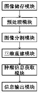
[0049] see figure 1 As shown, the present invention provides a three-dimensional tumor positioning system, including an image storage module, a preprocessing module, an image segmentation module, a three-dimensional reconstruction module, a tumor information acquisition module, and an information output module.

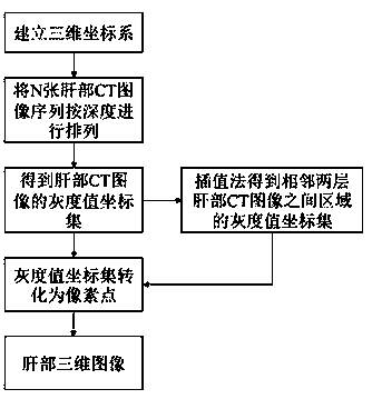
[0050] Taking liver tumors as an example, the image storage block is used to store two-dimensional CT images of different depths of the human liver and standard liver CT images to obtain liver CT image sequences and standa...


PUM


Abstract

The invention provides a tumor three-dimensional positioning system. The tumor three-dimensional positioning system comprises an image acquisition module, a preprocessing module, an image segmentationmodule, a three-dimensional reconstruction module, a tumor information acquisition module and an information output module. The tumor three-dimensional positioning system sequentially carries out preprocessing and image segmentation on the liver CT image to obtain a gray value matrix of the liver CT image sequence and a gray value matrix of an area where a tumor is located, performs interpolationoperation between layers of the liver two-dimensional CT image by utilizing the gray value information of two adjacent layers according to the difference of gray values of specific coordinates in thetwo layers of images and the gray relationship to obtain a new CT image, converts the gray value and the pixel points to obtain a three-dimensional image of the liver and the tumor, and finally, obtains tumor center coordinates and edge coordinates of a tumor and liver part interface through a tumor information obtaining module, thereby qualitatively and quantitatively presenting a tumor orientation. The tumor three-dimensional positioning method has the advantages of being high in precision and convenient to observe, diagnose, treat and position.

Description

technical field [0001] The invention belongs to the field of medical technology, in particular to a three-dimensional positioning system for tumors. Background technique [0002] As one of the diseases with the highest morbidity and mortality in the world, liver cancer seriously threatens people's health and life. At present, impact examination has become one of the most direct and effective ways to identify tumors. In the early diagnosis of tumors, multi-slice spiral CT can clearly display the lesion characteristics in the cross-section, sagittal plane and coronal plane through reconstruction technology. In the mid-term diagnosis, spiral CT Diagnosis combined with surface masking and multi-planar reconstruction can clearly display tumor location, internal structure, edge features, blood supply, degree of invasion of surrounding tissues, and changes of surrounding tissues, with high diagnostic accuracy. However, CT images can only provide two-dimensional images of the insid...

Claims


Application Information

Patent Timeline
IPC IPC(8): G06T7/70G06T7/12G06T7/30G06T7/13
CPCG06T7/12G06T7/13G06T7/30G06T7/70G06T2207/10012G06T2207/10081G06T2207/30056G06T2207/30096
Inventor 祝守芳左丙丽李金鹏
Owner 上海聚慕医疗器械有限公司
Learn More