
Heart magnetic resonance image analysis and cardiomyopathy prediction method and device

A magnetic resonance and cardiomyopathy technology, applied in the field of image processing, can solve problems such as low accuracy, lack of verification indicators, and inability to realize full automation, and achieve high accuracy

Inactive Publication Date: 2020-02-07
BEIJING ANDE YIZHI TECH CO LTD
View PDF5 Cites 12 Cited by
  • Summary
  • Abstract
  • Description
  • Claims
  • Application Information

AI Technical Summary

Problems solved by technology

However, conventional cardiac magnetic resonance image analysis cannot be fully automated, requiring a lot of manual operations, low efficiency, and errors caused by human factors. At the same time, there is a lack of indicators for verification, and the accuracy rate is low

Method used


Image

  • Heart magnetic resonance image analysis and cardiomyopathy prediction method and device
  • Heart magnetic resonance image analysis and cardiomyopathy prediction method and device
  • Heart magnetic resonance image analysis and cardiomyopathy prediction method and device

Examples


Embodiment Construction

[0045] Various exemplary embodiments, features, and aspects of the present disclosure will be described in detail below with reference to the accompanying drawings. The same reference numbers in the figures indicate functionally identical or similar elements. While various aspects of the embodiments are shown in drawings, the drawings are not necessarily drawn to scale unless specifically indicated.

[0046] The word "exemplary" is used exclusively herein to mean "serving as an example, embodiment, or illustration." Any embodiment described herein as "exemplary" is not necessarily to be construed as superior or better than other embodiments.

[0047] In addition, in order to better illustrate the present disclosure, numerous specific details are given in the following specific implementation manners. It will be understood by those skilled in the art that the present disclosure may be practiced without some of the specific details. In some instances, methods, means, componen...


PUM


Abstract

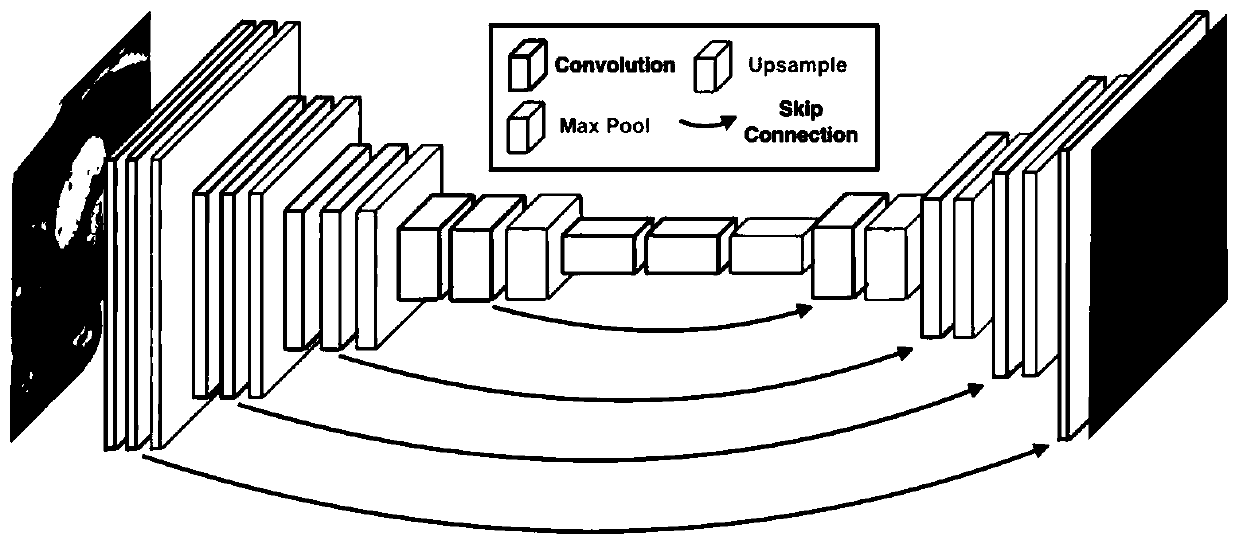
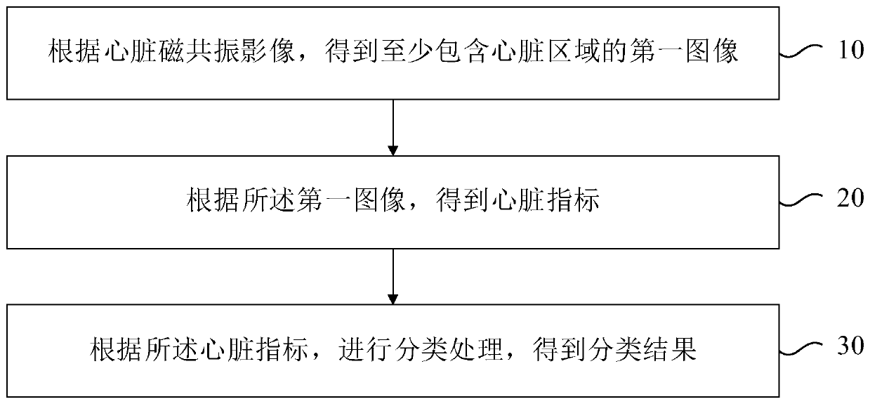
The invention relates to a heart magnetic resonance image analysis and cardiomyopathy prediction method and device, and the method carries out the processing through a trained neural network, and theprocessing at least comprises the steps: obtaining a first image at least comprising a heart region according to a heart magnetic resonance image; obtaining a heart index according to the first image;and performing classification processing according to the heart indexes to obtain a classification result. In the embodiment of the invention, the artificial intelligence analysis technology of the heart magnetic resonance image is adopted, the full automation of image analysis can be realized through the trained neural network, the analysis of the heart magnetic resonance image can be quickly completed, and higher accuracy can be achieved.

Description

technical field [0001] The present disclosure relates to the field of image processing, and in particular to a method and device for cardiac magnetic resonance image analysis and cardiomyopathy prediction. Background technique [0002] Cardiac magnetic resonance imaging is the gold standard for measuring the overall function of the heart, especially the left ventricular function, and diagnosing myocardial diseases. However, conventional cardiac magnetic resonance image analysis cannot be fully automated, requires a lot of manual operations, is inefficient, and has errors caused by human factors. At the same time, there is a lack of indicators for verification, and the accuracy rate is low. Contents of the invention [0003] In view of this, the present disclosure proposes a method and device for cardiac magnetic resonance image analysis and cardiomyopathy prediction. [0004] According to one aspect of the present disclosure, a method for cardiac magnetic resonance image ...

Claims


Application Information

Patent Timeline
IPC IPC(8): G06T7/00G06T7/11G06N3/08G06N3/04G06K9/62G16H30/20G16H70/00
CPCG06T7/0012G06T7/11G06N3/08G16H70/00G16H30/20G06T2207/10088G06T2207/30048G06N3/045G06F18/24
Inventor 张培芳罗志鹏廖宝鑫吴振洲
Owner BEIJING ANDE YIZHI TECH CO LTD
Learn More