
A method and device for dissecting artery segmentation

An arterial and dissection technology, applied in the field of medical image processing, can solve the problems of large data size, poor segmentation robustness of dissection model, and inability to identify true and false cavities individually, so as to improve segmentation efficiency, reduce image data, and maintain accuracy.

Active Publication Date: 2022-07-26
BEIJING INSTITUTE OF TECHNOLOGYGY
View PDF6 Cites 0 Cited by
  • Summary
  • Abstract
  • Description
  • Claims
  • Application Information

AI Technical Summary

Problems solved by technology

The existing traditional segmentation methods are less robust to the segmentation of sandwich models, and cannot identify true and false lumens alone. Moreover, due to the large size of CTA data, segmentation, segmentation accuracy, and 3D reconstruction efficiency need to be improved urgently.

Method used


Image

  • A method and device for dissecting artery segmentation
  • A method and device for dissecting artery segmentation
  • A method and device for dissecting artery segmentation

Examples


Embodiment Construction

[0065] In order to make the purposes, technical solutions and advantages of the embodiments of the present application clearer, the technical solutions in the embodiments of the present application will be clearly and completely described below with reference to the drawings in the embodiments of the present application. Obviously, the described embodiments are only It is a part of the embodiments of the present application, but not all of the embodiments. The components of the embodiments of the present application generally described and illustrated in the drawings herein may be arranged and designed in a variety of different configurations. Thus, the following detailed description of the embodiments of the application provided in the accompanying drawings is not intended to limit the scope of the application as claimed, but is merely representative of selected embodiments of the application. Based on the embodiments of the present application, every other embodiment obtaine...


PUM


Abstract

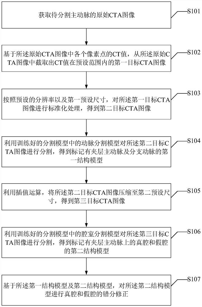
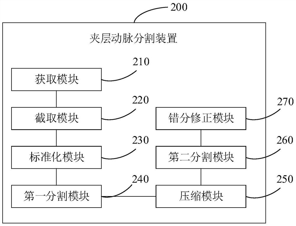
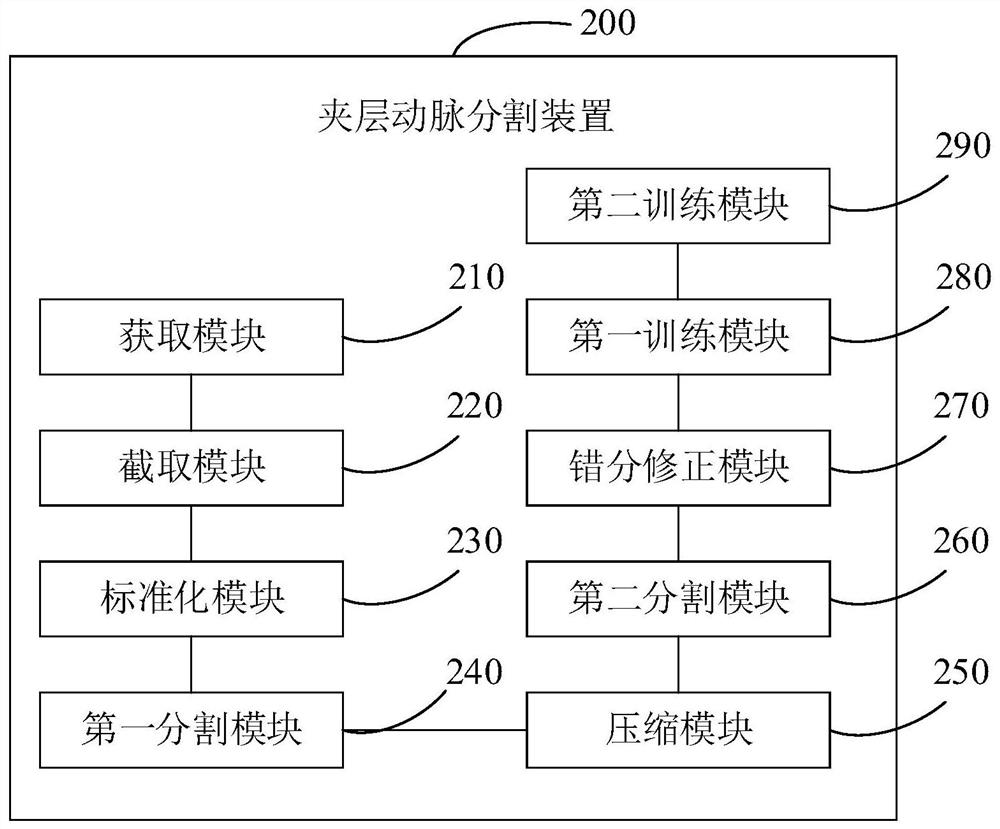
The present application provides a method and device for segmenting a dissecting artery, by acquiring an original CTA image of the aorta to be segmented; preprocessing the original CTA image to obtain a second target CTA image; using the artery segmentation model in the trained segmentation model and the chamber segmentation model, segment the second target CTA image to obtain a first structural model marked with the dissection aorta and branch arteries and a second structural model marked with the true lumen and false lumen on the dissection aorta; The first structural model and the second structural model are used to correct the misclassification of the true cavity and the false cavity on the second structural model. The present application standardizes the original CTA image, and uses the artery segmentation model and the chamber segmentation model to segment the standardized CTA image, which greatly reduces the image data that needs to be processed, effectively improves the efficiency of dissecting artery segmentation, and improves the the segmentation accuracy.

Description

technical field [0001] The present application relates to the technical field of medical image processing, and in particular, to a method and device for segmenting a dissecting artery. Background technique [0002] Aortic dissection (aortic dissection) is a very dangerous aortic disease, which is mainly characterized by the rupture of the intima of the aorta of the patient. , two cavities, the true cavity and the false cavity, are forcibly generated under the impact of blood, which is an extremely dangerous disease and requires timely diagnosis and treatment. Computed tomography angiography (CTA, CT angiography) is the most commonly used method for clinical diagnosis and observation of aortic dissection. Doctors usually analyze the condition based on CT scan tomography, which is not conducive to doctors' acquisition. Accurate 3D morphological features of dissecting aorta, such as determining the size and position of the tear opening, measuring the diameter of the aorta and ...

Claims


Application Information

Patent Timeline
Patent Type & Authority Patents(China)
IPC IPC(8): G06T7/11G06T7/194G06N3/04G06N3/08
CPCG06T7/11G06T7/194G06N3/08G06T2207/10081G06T2207/20081G06T2207/20084G06T2207/30101G06N3/045
Inventor 陈端端张栩阳李振锋梅玉倩梁世超石悦
Owner BEIJING INSTITUTE OF TECHNOLOGYGY
Learn More