Superfine thoracoscope

A thoracoscopic and mirror body technology, applied in the field of thoracoscopy, can solve the problems of obvious trauma and bleeding risks, achieve the effect of solving trauma and bleeding risks, less channel damage, and increasing compliance

Inactive Publication Date: 2020-02-25
上海市闵行区中心医院
View PDF8 Cites 0 Cited by
  • Summary
  • Abstract
  • Description
  • Claims
  • Application Information

AI Technical Summary

Problems solved by technology

[0003] The purpose of the present invention is to provide an ultra-fine thoracoscope to solve the problems that the existing medical thoracoscopes in the above background technology easily cause obvious trauma and bleeding risks

Method used


Image

  • Superfine thoracoscope
  • Superfine thoracoscope
  • Superfine thoracoscope

Examples


Embodiment 1

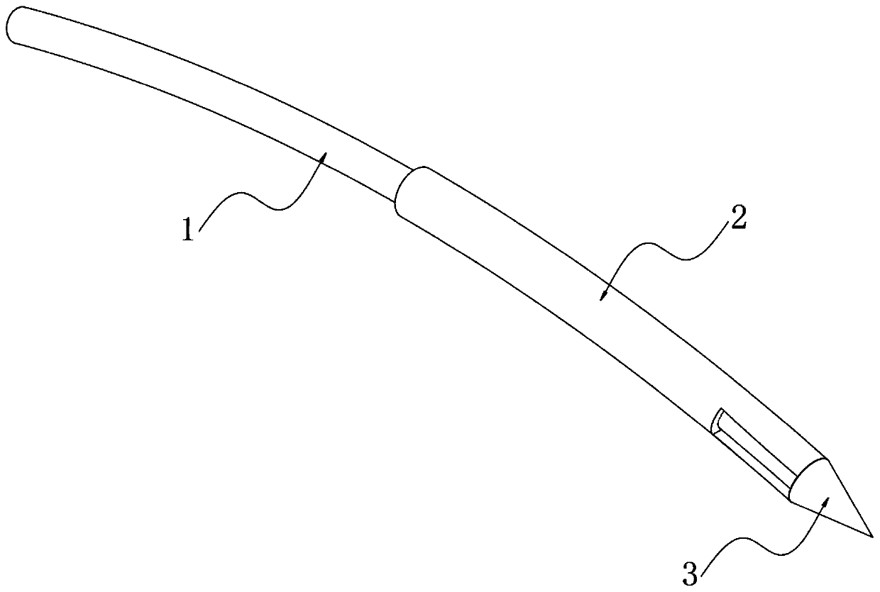
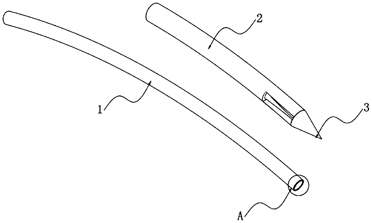
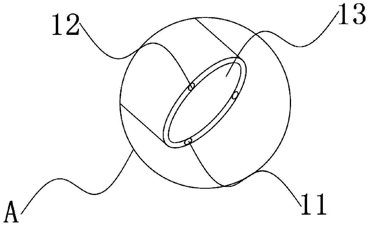
[0027] An ultra-thin thoracoscope, such as Figure 1 to Figure 4 As shown, a mirror body 1 is included, a sleeve 2 is sheathed on the mirror body 1 , and a blunt puncture needle 3 is embedded at the end of the mirror body 1 . A biopsy suction channel 11 is opened inside the mirror body 1 , and two symmetrical glass fiber light source bundles 12 are embedded inside the mirror body 1 , and an image acquisition hole 13 is opened on one side of the biopsy suction channel 11 . Two rectangular grooves 21 are formed symmetrically near the blunt puncture needle 3 on the outer peripheral wall of the cannula 2 .

[0028] Further, the diameter of the mirror body 1 is 4 mm, and the diameter of the sleeve 2 is 5 mm, which is convenient for the sleeve 2 to be sleeved on the mirror body 1, and facilitates the sleeve 2 to drive the mirror body 1 to puncture into the patient's body without causing serious complications. Large wounds.

[0029] In addition, the mirror body 1 is made of stainle...

Embodiment 2

[0035] As the second embodiment in this embodiment, please refer to Figure 5 to Figure 8 , which is different from Embodiment 1 in that: the mirror body 1 is provided with a fixing mechanism 4 . Specifically, the fixing mechanism 4 includes a fixed disk 41, a plurality of suction cups 43 are closely bonded to each other at equal intervals in a ring on the right side of the fixed disk 41, and a through hole 42 is provided on the mirror body 1, and the left side of the fixed disk 41 A protruding tube 44 is tightly bonded at the center, and the outer wall of the protruding tube 44 is closely bonded with a limit ring 442 coaxially close to the left end. The protruding tube 44 is provided with a plurality of strip-shaped grooves 441 arranged at equal intervals in a ring, and the strips The shaped groove 441 runs through the limit ring 442 , the rubber ring 45 is sleeved on the convex tube 44 , and the mirror body 1 passes through the through hole 42 and the convex tube 44 in turn....


PUM


Abstract

The invention relates to the technical field of thoracoscopes, in particular to a superfine thoracoscope, which comprises a thoracoscope body, the thoracoscope body is sleeved with a sleeve, and a blunt puncture needle is embedded in the end of the thoracoscope body. A biopsy suction hole channel is formed in the endoscope body, two symmetrical glass fiber light source beams are further embedded in the endoscope body, and an image collecting hole is further formed in one side of the biopsy suction hole channel. According to the superfine thoracoscope, the blunt puncture needle with the sleevepenetrates into the thoracic cavity, intercostal blood vessels and nerves are not prone to being damaged, and the risk of bleeding is avoided; meanwhile, the mirror bodies with two radians are arranged, so that the thoracic wall focus and biopsy are convenient to observe, and far and near focuses are convenient to detect; in addition, compared with a channel formed by traditional puncture, a puncture channel formed by the arrangement is small in damage, suture is not needed after operation, the compliance of a patient is improved, and the problem that an existing thoracoscope for the internalmedicine department is prone to causing obvious trauma and bleeding risks is solved.

Description

technical field [0001] The invention relates to the technical field of thoracoscopy, in particular to an ultra-thin thoracoscope. Background technique [0002] The current medical thoracoscopes are divided into soft and hard, but their diameters are more than 8mm. To enter the chest cavity, an incision of about 1cm is required in the skin, and the subcutaneous and muscle tissue are separated, which not only causes obvious trauma, but also has the risk of bleeding; Due to trauma and risk, some patients are unwilling to undergo medical thoracoscopy, which brings inconvenience to diagnosis and treatment. In view of this, we propose an ultra-thin thoracoscopy. Contents of the invention [0003] The purpose of the present invention is to provide an ultra-thin thoracoscope to solve the problems that the existing medical thoracoscopes easily cause obvious trauma and bleeding risks in the background art. [0004] To achieve the above object, the present invention provides the fol...

Claims


Application Information

Patent Timeline
Patent Type & Authority Applications(China)
IPC IPC(8): A61B1/00A61B1/07A61B1/04A61B17/34
CPCA61B1/00071A61B1/04A61B1/07A61B17/3417A61B17/3478
Inventor 高习文
Owner 上海市闵行区中心医院
Why Eureka
Try Eureka