
Optical coherence tomography method

A technology of optical coherence tomography and imaging method, which is applied in the directions of diagnosis, medical science, and diagnostic signal processing, etc. It can solve the problems of slow speed and restricted application, and achieve the effect of reducing complexity and improving the depth of tomographic imaging

Active Publication Date: 2020-03-17
BEIJING INFORMATION SCI & TECH UNIV
View PDF6 Cites 6 Cited by
  • Summary
  • Abstract
  • Description
  • Claims
  • Application Information

AI Technical Summary

Problems solved by technology

However, since such technical solutions are all based on the time-domain OCT (TD-OCT) system, the measurement time for the two-dimensional transmission matrix of a single target layer generally takes about 6-7 minutes, which is very slow, which restricts its application in biological Applications in Tissue Imaging

Method used


Image

  • Optical coherence tomography method
  • Optical coherence tomography method
  • Optical coherence tomography method

Examples


Embodiment Construction

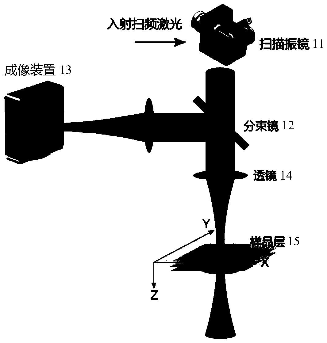
[0047] In order to make the technical solutions and advantages of the present invention clearer, the present invention will be further described in detail below in conjunction with the accompanying drawings and specific embodiments.

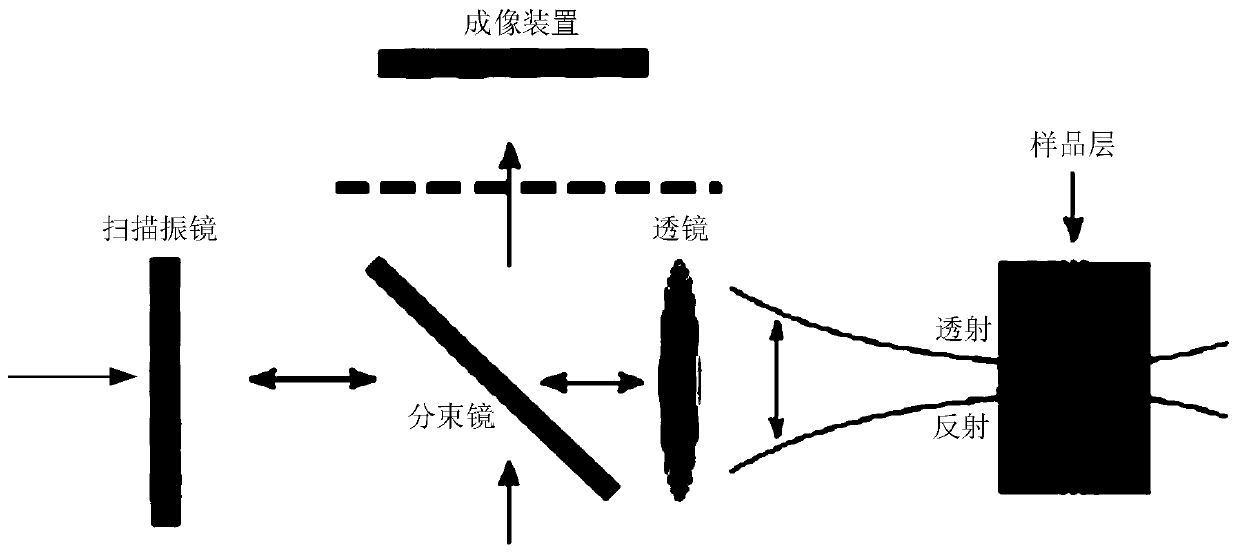
[0048]The OCT system in the prior art usually uses an optical fiber (pinhole) as the medium at the signal receiving end, and the detector records the interference echo generated during the scanning process, and then generates a two-dimensional or three-dimensional tomographic image according to the echo signal. Similar to a pinhole in a confocal microscope, the optical fiber receiving end of an OCT system in the prior art can filter out optical echoes in regions other than the focal region, and suppress background echoes and scattering noise from non-focused regions.

[0049] However, just like the physical phenomenon that a stone stirs up waves, the two-dimensional transverse light field distribution in the adjacent area of ​​the focused scanning...


PUM


Abstract

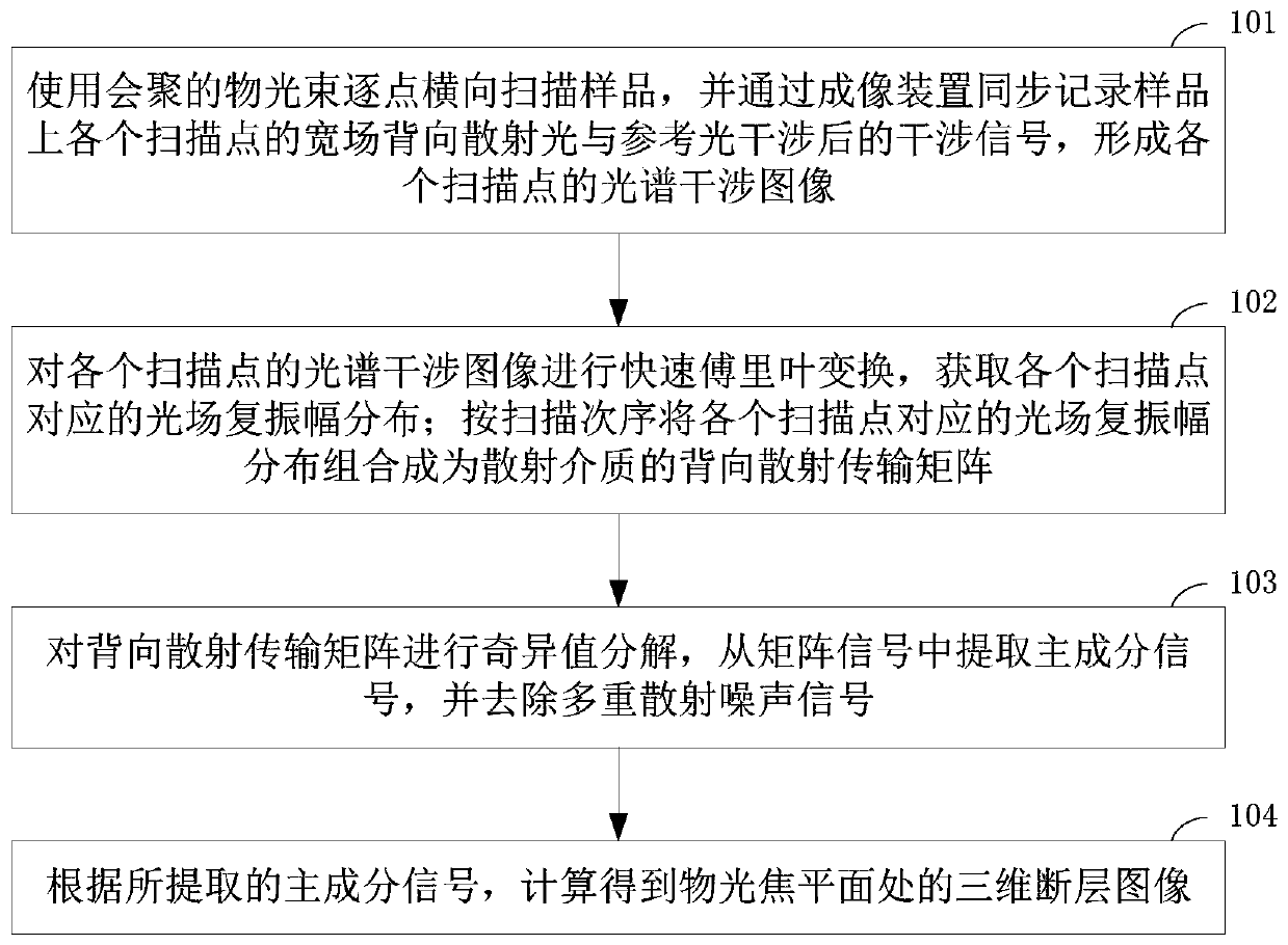
The invention provides an optical coherence tomography method. The method comprises the following steps: transversely scanning a sample point by point by using a converged object light beam, synchronously recording an interference signal after interference between wide-field backscattered light of each scanning point on the sample and reference light through an imaging device to form a spectral interference image of each scanning point, performing fast Fourier transform on the spectral interference image of each scanning point to obtain optical field complex amplitude distribution corresponding to each scanning point, combining the optical field complex amplitude distribution corresponding to the scanning points in a scanning order into a back transmission matrix of the scattering medium,carrying out singular value decomposition on the back transmission matrix, extracting principal component signals from matrix signals, removing multiple scattering noise signals and calculating a three-dimensional tomographic image at the object focal plane according to the extracted principal component signal. According to the method, tissue deep imaging can be realized and the imaging depth andthe imaging speed are improved.

Description

technical field [0001] The present application relates to the technical field of optical imaging, in particular to an optical coherence tomography method. Background technique [0002] Optical coherence tomography (OCT, Optical Coherence Tomography) technology uses the interference principle of a wide-spectrum light source to extract the layered information of the imaged tissue by detecting the interference signal between the backscattered light of the sample and the reference light. Because of its non-invasive, non-contact characteristics and tomographic imaging capabilities, OCT technology is widely used in ophthalmology, vascular endoscopy and other clinical medicine and pharmacology. [0003] Compared with the imaging depth of a confocal scanning microscope of about 100 microns and a two-photon microscope of about a few hundred microns to 1 mm, the traditional OCT system can image in human soft tissues at a depth of about 1-2 mm. Imaging capabilities and micron-level im...

Claims


Application Information

Patent Timeline
IPC IPC(8): A61B3/10A61B3/12A61B3/14A61B5/00G01N21/45
CPCA61B3/102A61B3/1225A61B3/14A61B5/0066A61B5/72A61B5/7203G01N21/45
Inventor 杨强祝连庆朱疆樊凡端木正
Owner BEIJING INFORMATION SCI & TECH UNIV
Learn More