System for identifying and positioning pancreatic neuroendocrine tumors under ultrasonic endoscope

A neuroendocrine and pancreas technology, applied in ultrasound/sonic/infrasonic Permian technology, ultrasonic/sonic/infrasonic diagnosis, ultrasonic/sonic/infrasound image/data processing, etc. , misdiagnosis and missed diagnosis, etc., to improve the detection rate, reduce missed diagnosis, and improve reliability.

Active Publication Date: 2020-04-10
SHANDONG UNIV QILU HOSPITAL +1
View PDF5 Cites 5 Cited by
  • Summary
  • Abstract
  • Description
  • Claims
  • Application Information

AI Technical Summary

Problems solved by technology

However, because the endoscopic ultrasonographic images of PNET lesions have a very high similarity to the images of the normal pancreatic background, and there will be a large number of endoscopic ultrasonographic images during the continuous scanning of endoscopic ultrasonography, the PNET should be examined during the examination process. It is difficult to identify and locate, and requires the examiner to have rich experience in endoscopic ultrasonography, and there is a certain degree of misdiagnosis and missed diagnosis

Method used


Image

  • System for identifying and positioning pancreatic neuroendocrine tumors under ultrasonic endoscope

Examples


Embodiment 1

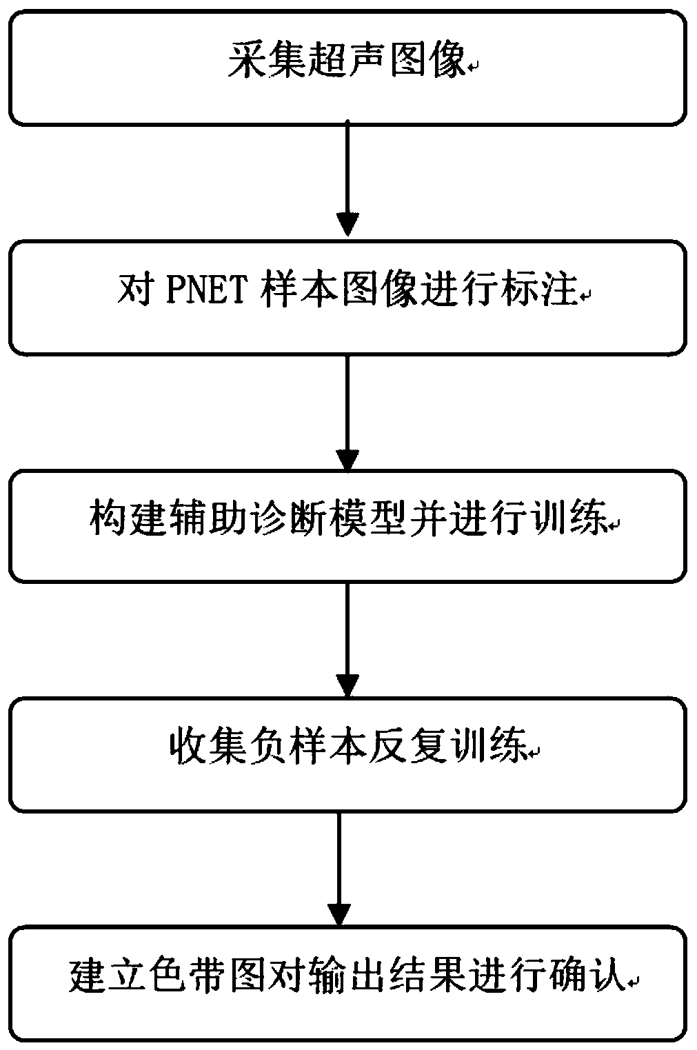
[0035] In one or more embodiments, a system for identifying and locating pancreatic neuroendocrine tumors under endoscopic ultrasonography is disclosed, comprising:

[0036] The image acquisition module is connected to the endoscope host through the acquisition card to obtain each frame of image information collected by the endoscope host; construct a sample set by manually selecting a single frame of endoscope images with PNET lesions;

[0037] An image preprocessing module configured to preprocess the collected image information;

[0038] Since the images with PNET lesions are collected in a single sheet under endoscopic ultrasonography in clinic, it is necessary to remove the patient's private data in the images. In order to reduce the amount of calculation, it is necessary to remove the black frame and only keep the colored digestive tract area.

[0039] Through the black edge algorithm processing, zoom processing and normalization processing, each frame of the image is p...

Embodiment 2

[0072] In one or more embodiments, a terminal device is disclosed, which includes a processor and a computer-readable storage medium, the processor is used to implement instructions; the computer-readable storage medium is used to store multiple instructions, and the instructions are suitable for to be loaded and executed by the processor as figure 1 The described process:

[0073] Connect the endoscope host through the acquisition card to obtain each frame of image information collected by the endoscope host; select a single frame of endoscopic images with PNET lesions to construct a sample set;

[0074] Using a multi-target labeling tool to label the pancreatic neuroendocrine tumor region in the image of the sample set; at the same time, generating labeling text information corresponding to the labeling position; the labeling area and the labeling text information corresponding to the area constitute a training set;

[0075] Construct an auxiliary diagnosis model, and after...


PUM


Abstract

The invention discloses a system for identifying and positioning pancreatic neuroendocrine tumors under an ultrasonic endoscope. The system comprises an image acquisition module, a training set manufacturing module, an auxiliary diagnosis module and a joint judgment module; the training set manufacturing module is configured to mark pancreatic neuroendocrine tumor regions in a sample set image byusing a multi-target marking tool; the auxiliary diagnosis module is configured to construct an auxiliary diagnosis model, and is used for performing identification of pancreatic neuroendocrine tumorlesion regions on an input pretreated image after optimization training of a training set; and the joint judgment module is configured to display an output result in a form of a color bar pattern, andis used for judging accuracy of the identification result of the pancreatic neuroendocrine tumor lesion regions. Through intelligent and automatic identification of pancreatic neuroendocrine tumors (PNET) under an ultrasonic endoscope, the PNET can be accurately identified and positioned in a large number of generated ultrasonic endoscope pictures in an ultrasonic endoscope examination process, so that the detection rate of the PNET is improved, and missed diagnosis is reduced.

Description

technical field [0001] The invention relates to the technical field of intelligent auxiliary diagnosis of tumors, in particular to a system for identifying and locating pancreatic neuroendocrine tumors under an ultrasonic endoscope. Background technique [0002] The statements in this section merely provide background information related to the present invention and do not necessarily constitute prior art. [0003] At present, the identification and location of pancreatic neuroendocrine tumors (PNET) under endoscopic ultrasonography still depend on the operator of endoscopic ultrasonography. PNET. However, because the endoscopic ultrasonographic images of PNET lesions have a very high similarity to the images of the normal pancreatic background, and there will be a large number of endoscopic ultrasonographic images during the continuous scanning of endoscopic ultrasonography, the PNET should be examined during the examination process. Recognition and positioning are diffic...

Claims


Application Information

Patent Timeline
Patent Type & Authority Applications(China)
IPC IPC(8): A61B8/08A61B8/12A61B8/00
CPCA61B8/12A61B8/463A61B8/5223
Inventor 李真戚庆庆冯建左秀丽李延青杨晓云邵学军季锐
Owner SHANDONG UNIV QILU HOSPITAL
Why Eureka
Try Eureka