Lower digestive tract endoscope image recognition method and system

An image recognition and digestive tract technology, which is applied in the field of digestive tract endoscope image recognition, can solve the problems of affecting the quality of lower digestive tract endoscopy, the inability to accurately judge the lesion site, and the inability to accurately determine, so as to facilitate treatment and follow-up, The effect of improving recognition accuracy and improving precision

Active Publication Date: 2020-04-10
SHANDONG UNIV QILU HOSPITAL +1
View PDF10 Cites 3 Cited by
  • Summary
  • Abstract
  • Description
  • Claims
  • Application Information

AI Technical Summary

Problems solved by technology

The inventors found that at present, most of the endoscopic examinations of the lower gastrointestinal tract are observed by human eyes, and the lesion site cannot be accurately judged.
For example, multiple polyps in the lower gastrointestinal tract were found by visual observation, but it was not possible to accurately determine whether they were located in the ascending colon, transverse colon, descending colon, or sigmoid colon;
[0004] In addition, some parts may be missed during the lower gastrointestinal endoscopy, for example, the image quality of a certain part is poor due to the slipping of the mirror body or poor filling of the intestinal cavity, which affects the quality of the lower gastrointestinal endoscopy, resulting in Possibility of missed diagnosis

Method used


Image

  • Lower digestive tract endoscope image recognition method and system
  • Lower digestive tract endoscope image recognition method and system
  • Lower digestive tract endoscope image recognition method and system

Examples


Embodiment 1

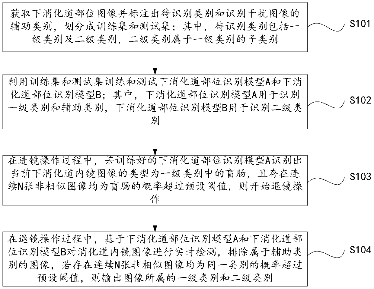
[0030] figure 1 A flow chart of a lower gastrointestinal endoscope image recognition method in this embodiment is given.

[0031] Combine below figure 1 To describe in detail the specific implementation process of the lower gastrointestinal endoscope image recognition method of this embodiment:

[0032] Such as figure 1 As shown, a kind of lower gastrointestinal endoscope image recognition method of the present embodiment at least includes:

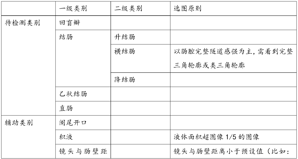
[0033] Step S101: Obtain the image of the lower gastrointestinal tract, mark the category to be recognized and the auxiliary category of the recognition interference image, and divide it into a training set and a test set; wherein, the category to be recognized includes a first-level category and a second-level category, and the second-level category belongs to the first-level category. subcategory of the level category.

[0034] In the specific implementation, the primary category includes the ileocecal valve, colon, sigmoid colon, a...

Embodiment 2

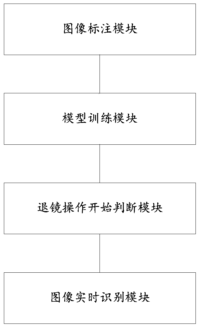
[0070] figure 2 A schematic structural diagram of a lower gastrointestinal endoscope image recognition system of this embodiment is given.

[0071] Combine below figure 2 To describe in detail the structural composition of the lower gastrointestinal endoscope image recognition system of this embodiment:

[0072] Such as figure 2 As shown, a kind of lower gastrointestinal endoscope image recognition system of the present embodiment at least includes:

[0073] (1) Image labeling module, which is used to obtain the image of the lower digestive tract and mark the auxiliary category of the category to be identified and the recognition interference image, and is divided into a training set and a test set; wherein, the category to be identified includes a first-level category and a second-level category Category, the secondary category is a subcategory of the primary category.

[0074] In the specific implementation, the primary category includes the ileocecal valve, colon, si...

Embodiment 3

[0110] This embodiment provides a computer-readable storage medium on which a computer program is stored, and it is characterized in that, when the program is executed by a processor, the following figure 1 The steps in the image recognition method for lower gastrointestinal endoscopy are shown.

[0111] In this embodiment, the image of the lower digestive tract is obtained and the category to be identified and the auxiliary category for identifying the interference image are marked; wherein, the category to be identified includes a first-level category and a second-level category, and the second-level category belongs to a subcategory of the first-level category; The lower gastrointestinal part images of the auxiliary category and the lower gastrointestinal part images of the first-level category are used to train the lower gastrointestinal part recognition model A, which reduces the impact of blurred images, out-of-focus images, reflection artifacts, residual debris, and effu...


PUM


Abstract

The invention provides a lower digestive tract endoscope image recognition method and system. The method comprises the following steps of: acquiring a lower digestive tract part image, marking a to-be-identified category and an auxiliary category for identifying an interference image, and dividing into a training set and a test set, training and testing a lower digestive tract part recognition model A and a lower digestive tract part recognition model B by using the training set and the test set, in the endoscope entering operation process, if the lower digestive tract part recognition model Arecognizes that the type of the current lower digestive tract endoscope image is the cecum in the first-level category and the probability that the continuous N non-similar images are all the cecum exceeds a preset threshold value, starting endoscope retreating operation, N being a positive integer greater than or equal to 3, and in the retreating operation process, detecting the digestive tractendoscope image in real time based on the lower digestive tract part recognition model A and the lower digestive tract part recognition model B, excluding the image belonging to the auxiliary category, and outputting the first-level category and the second-level category to which the image belongs.

Description

technical field [0001] The disclosure belongs to the field of gastrointestinal endoscope image recognition, in particular to a lower gastrointestinal endoscope image recognition method and system. Background technique [0002] The statements in this section merely provide background information related to the present disclosure and do not necessarily constitute prior art. [0003] In lower gastrointestinal endoscopy, it is often necessary to determine where a lesion is located in the colorectum. The inventors found that at present, most of the endoscopic examinations of the lower gastrointestinal tract are observed by human eyes, and the lesion site cannot be accurately judged. For example, multiple polyps in the lower gastrointestinal tract were found by visual observation, but it was not possible to accurately determine whether they were located in the ascending colon, transverse colon, descending colon, or sigmoid colon; [0004] In addition, some parts may be missed du...

Claims


Application Information

Patent Timeline
IPC IPC(8): G06K9/62
CPCG06V2201/03G06F18/2415G06F18/214
Inventor 李延青李广超冯建左秀丽杨晓云邵学军赖永航辛伟李真
Owner SHANDONG UNIV QILU HOSPITAL
Why Eureka
Try Eureka