
CT image liver artery segmentation method and system based on deep learning

A CT image and deep learning technology, applied in the field of medical image processing and artificial intelligence, can solve the problems of difficult expansion and poor adaptability, and achieve the effect of improving segmentation efficiency, good robustness, strong representation ability and generalization ability

Inactive Publication Date: 2020-04-10
天津精诊医疗科技有限公司
View PDF6 Cites 2 Cited by
  • Summary
  • Abstract
  • Description
  • Claims
  • Application Information

AI Technical Summary

Problems solved by technology

[0005] In order to solve the problems existing in the existing hepatic artery region segmentation methods, such as humanized parameter setting, poor adaptability and difficult expansion caused by fixed parameters, the present invention provides a CT image hepatic artery segmentation method based on deep learning, including:

Method used


Image

  • CT image liver artery segmentation method and system based on deep learning
  • CT image liver artery segmentation method and system based on deep learning
  • CT image liver artery segmentation method and system based on deep learning

Examples


Embodiment Construction

[0051] The technical solutions of the present invention will be further described below in conjunction with the accompanying drawings and embodiments.

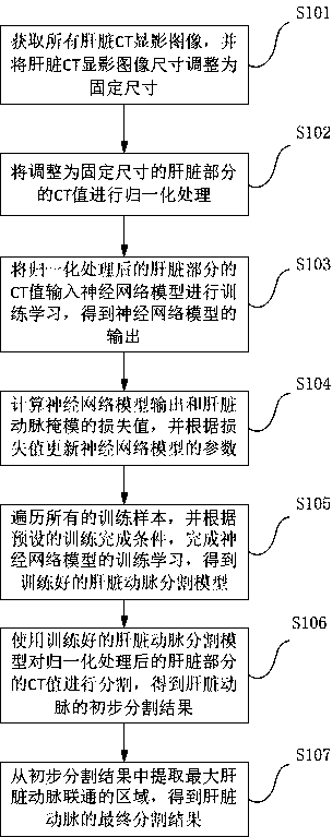
[0052] see figure 1 , the deep learning-based CT image hepatic artery segmentation method provided by the embodiment of the present invention specifically includes the following steps:

[0053]Step S101 , acquiring all liver CT developed images, and adjusting the size of the liver CT developed images to a fixed size.

[0054] Usually abdominal CT images contain hundreds of slices, and there are visualization of the liver and other organs at the same time. Since the segmentation of the hepatic artery only depends on the difference in imaging between intrahepatic tissue and arteries inside the liver, liver imaging needs to be extracted from hundreds of slices for hepatic artery segmentation. The specific process is as follows:

[0055] 1) Cut out the liver visualization in the whole abdominal CT image to exclude the influence ...


PUM


Abstract

The invention discloses a CT image liver artery segmentation method and system based on deep learning, and belongs to the technical field of medical image processing and artificial intelligence. The method comprises the steps that all liver CT development images are acquired, and the image size is adjusted to a fixed size; normalizing the CT value of the liver part, and inputting the normalized CTvalue into a neural network model; calculating the output of the neural network model and the loss value of a liver artery mask, and updating the parameters of the neural network model according to the loss value; traversing all the training samples, and completing training learning of the neural network model to obtain a liver artery segmentation model; and according to the liver artery segmentation model, segmenting the normalized CT value of the liver part to obtain a segmentation result of the liver artery. The system comprises an acquisition module, an adjustment module, a normalizationinput module, a calculation updating module, a training module and a segmentation module. According to the method, the actual condition of the hepatic artery can be accurately obtained, and no human participation is needed in the segmentation process.

Description

technical field [0001] The present invention relates to the technical fields of medical image processing and artificial intelligence, in particular to a method and system for segmenting liver arteries in CT images based on deep learning. Background technique [0002] Medical imaging has a variety of image modalities, including MR (Magnetic Resonance, magnetic resonance), CT (Computed Tomography, computerized tomography), PET (Positron Emission Computed Tomography, positron emission computed tomography), ultrasound imaging, etc. . Imaging can obtain images that reflect the physiological and physical properties of the human body in two-dimensional and three-dimensional regions. Patients with liver disease need to take enhanced CT before surgery. According to the different scanning time after the contrast agent enters the patient's body, enhanced CT can be divided into three-phase sequence images: arterial phase, portal vein phase, and scanning phase. By segmenting the develo...

Claims


Application Information

Patent Timeline
Patent Type & Authority Applications(China)
IPC IPC(8): G06T7/11G06T7/136G06N3/04
CPCG06T7/11G06T7/136G06T2207/10081G06T2207/20081G06T2207/30056G06T2207/30101G06N3/045
Inventor 赵威申建虎代笃伟王博张伟
Owner 天津精诊医疗科技有限公司
Learn More