Fluorescence-patch clamp-micropipette detection device

A detection device, patch clamp technology, applied in measurement devices, fluorescence/phosphorescence, material analysis through optical means, etc., can solve problems such as the inability to study the influence of membrane protein transmembrane signals

Active Publication Date: 2020-05-08
ZHEJIANG UNIV
View PDF12 Cites 4 Cited by
  • Summary
  • Abstract
  • Description
  • Claims
  • Application Information

AI Technical Summary

Problems solved by technology

[0005] In addition, fluorescence imaging technology is widely used in the study of transmembrane signal transduction of membrane proteins. Only the integration of patch clamp technology into micropipett

Method used


Image

  • Fluorescence-patch clamp-micropipette detection device
  • Fluorescence-patch clamp-micropipette detection device
  • Fluorescence-patch clamp-micropipette detection device

Examples


Embodiment Construction

[0027] The present invention will be further described below in conjunction with drawings and embodiments.

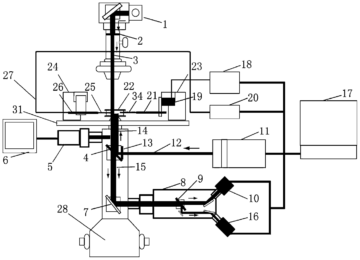
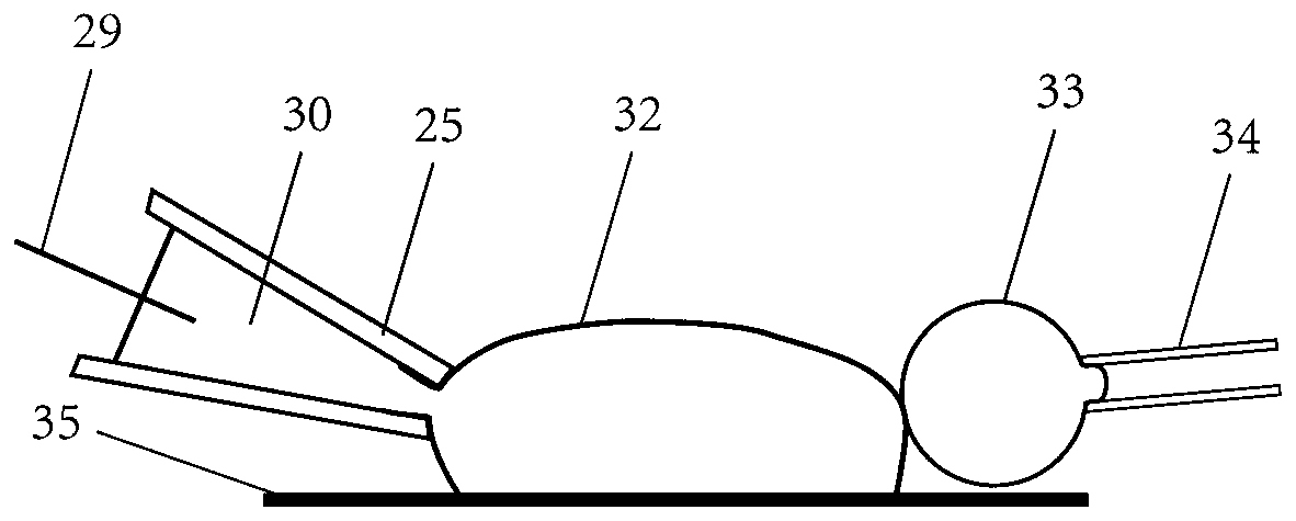
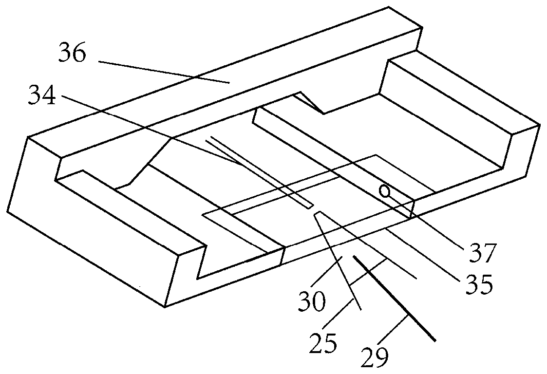
[0028] Such as figure 1 , shown in 2, the device of specific implementation comprises piezoelectric motion platform 19, micropipette holder 21, experiment chamber 22, first three-dimensional micromanipulator 23, second three-dimensional micromanipulator 24, glass electrode 25, Recording electrode 29, experimental platform 31, micropipette 34 and experimental cavity skeleton 36; experimental platform 31 is arranged with experimental cavity 22, first three-dimensional micromanipulator 23, second three-dimensional micromanipulator 24, patch clamp probe holder, etc. Such as image 3 As shown, the experimental cavity skeleton 36 is located at the center of the experimental platform 31, and the surface of the experimental cavity skeleton 36 is pasted with two parallel glass sheets up and down. The two glass sheets form the experimental cavity 22, and the two sides of the expe...


PUM


Abstract

The invention discloses a fluorescence-patch clamp-micropipette detection device. An experimental accommodating cavity, a three-dimensional micromanipulator and a patch clamp are arranged on an experiment platform, and two sides of the experimental accommodating cavity are hollowed out for a micropipette and a glass electrode to enter; fluorescent cells are contained in the experimental accommodating cavity, the micropipette sucks red blood cells, the fluorescent cells and the red blood cells are positioned in an extracellular fluid in the experimental accommodating cavity, and the glass electrode is filled with an electrode fluid; the patch clamp comprises a patch clamp amplifier, a patch clamp probe and a patch clamp probe bracket; full-spectrum light emitted from the fluorescence lightsource passes through a color filter to form fluorescence incident light, the fluorescence incident light enters the cells in the experimental accommodating cavity, and fluorescence of the cells returns to be received by means of a fluorescence camera. According to the fluorescence-patch clamp-micropipette detection device, the influence of the cell membrane potential change on the membrane protein interaction can be detected while a high signal-to-noise ratio fluorescence image is collected to research transmembrane signal transduction of the membrane protein, and the coupling relationship among the three spectrum phase information can be recorded while the fluorescence spectrum, the electrophysiological spectrum, the adhesion state spectrum and other three spectrum phase information aresynchronously recorded.

Description

technical field [0001] The invention relates to the integration of fluorescence imaging technology, patch clamp technology, and micropipette technology, and in particular to a detection device capable of studying membrane protein transmembrane signal transduction while detecting the regulation of membrane potential on the interaction between protein molecules. Background technique [0002] The membrane potential of the cell membrane is a key regulator of cell life activities. The membrane potential of neurons plays an important role in regulating the life activities of neurons, and is an important biophysical factor that dynamically regulates the structure and function of brain neural networks; in non-neuronal cells, the membrane potential of cells also regulates their proliferation , differentiation and other life activities. Patch clamp technology is an effective means to study membrane potential-related issues, and its whole-cell recording mode can realize accurate and r...

Claims


Application Information

Patent Timeline
IPC IPC(8): G01N21/64
CPCG01N21/6486
Inventor 陈伟安宸毅刘俊伟胡炜
Owner ZHEJIANG UNIV
Why Eureka
Try Eureka