Blood vessel segmentation method of medical image, computer equipment and readable storage medium

A technology of medical imaging and computer programs, which is applied in the field of image processing and can solve problems such as unsatisfactory auxiliary effects for film reading

Pending Publication Date: 2020-05-15
SHANGHAI UNITED IMAGING INTELLIGENT MEDICAL TECH CO LTD
View PDF14 Cites 16 Cited by
  • Summary
  • Abstract
  • Description
  • Claims
  • Application Information

AI Technical Summary

Problems solved by technology

[0005] Based on this, it is necessary to provide a medical imaging blood vessel segmentation method, computer equipment and readable storage medium for the problem that the blood vessel segmentation results obtained in the traditional technology are relatively general, and the effect of assisting doctors in film reading is not ideal.

Method used


Image

  • Blood vessel segmentation method of medical image, computer equipment and readable storage medium
  • Blood vessel segmentation method of medical image, computer equipment and readable storage medium
  • Blood vessel segmentation method of medical image, computer equipment and readable storage medium

Examples


Embodiment Construction

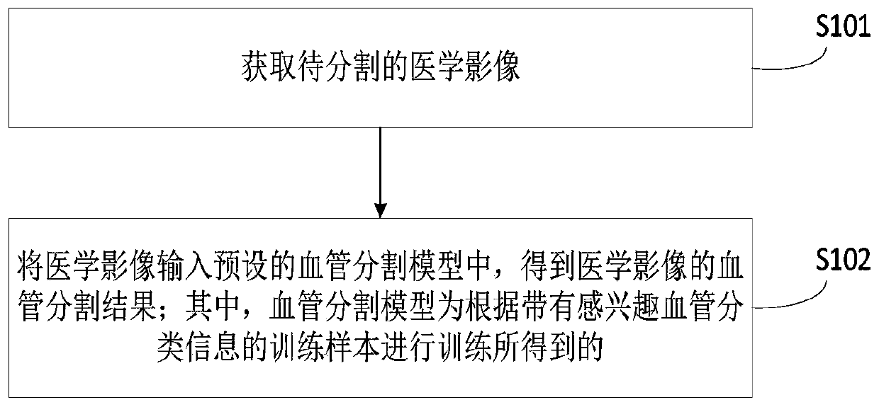
[0048] In order to make the purpose, technical solution and advantages of the present application clearer, the present application will be further described in detail below in conjunction with the accompanying drawings and embodiments. It should be understood that the specific embodiments described here are only used to explain the present application, and are not intended to limit the present application.

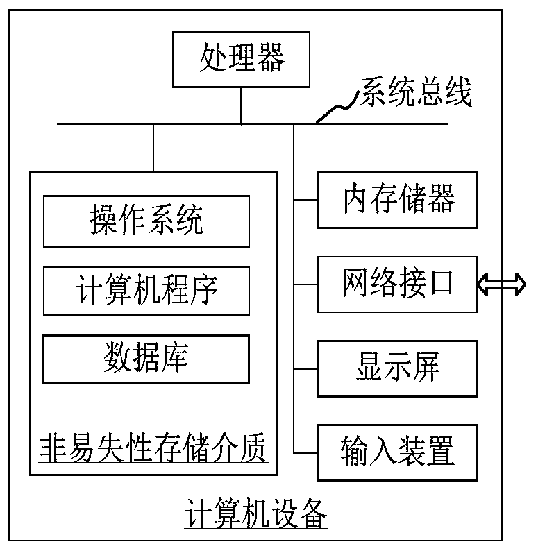
[0049] The blood vessel segmentation method for medical images provided in the embodiment of this application can be applied to such as figure 1computer equipment shown. The computer device includes a processor and a memory connected through a system bus, and a computer program is stored in the memory. When the processor executes the computer program, the steps of the following method embodiments can be performed. Optionally, the computer device may also include a network interface, a display screen and an input device. Wherein, the processor of the computer device is us...


PUM


Abstract

The invention relates to a blood vessel segmentation method of a medical image, computer equipment and a readable storage medium. The method comprises the steps of obtaining a to-be-segmented medicalimage; and inputting the medical image into a preset blood vessel segmentation model to obtain a blood vessel segmentation result of the medical image. The blood vessel segmentation model being obtained by training according to a training sample with interested blood vessel classification information. According to the blood vessel segmentation model obtained by training through the method, classification information of blood vessels (namely important blood vessels) which a doctor is interested in is considered, the blood vessel segmentation model can pay more attention to the segmentation process of the important blood vessels, the segmentation result of the important blood vessels in the obtained blood vessel segmentation result is prominent or obvious, and the doctor can be better assisted in film reading.

Description

technical field [0001] The present application relates to the technical field of image processing, in particular to a blood vessel segmentation method for medical images, computer equipment and a readable storage medium. Background technique [0002] Vascular analysis plays an important role in the clinical field, and vessel segmentation is an extremely important step in the vascular analysis process. Early blood vessel segmentation methods were mainly based on image analysis of angiographic images to obtain blood vessel segmentation results, but they were easily interfered by perivascular bone or plaque, which affected the measurement of the degree of vascular stenosis; The contrast-enhanced image is processed, but it requires the patient to remain fixed during the scan, and slight movement will lead to unsatisfactory results of the subtraction image. [0003] At present, deep learning has been gradually applied to the task of blood vessel segmentation. The supervised lear...

Claims


Application Information

Patent Timeline
Patent Type & Authority Applications(China)
IPC IPC(8): G06T7/10
CPCG06T7/10G06T2207/10081G06T2207/10088G06T2207/10104G06T2207/20081G06T2207/20084G06T2207/30101
Inventor 江鹏博石峰
Owner SHANGHAI UNITED IMAGING INTELLIGENT MEDICAL TECH CO LTD
Why Eureka
Try Eureka