Medical image lesion area segmentation method based on energy functional model of machine learning

An energy functional and medical image technology, applied in image analysis, image enhancement, image data processing, etc., to achieve the effect of retaining feature information, reliable accuracy, and guaranteed accuracy

Active Publication Date: 2020-05-22
LIAONING NORMAL UNIVERSITY
View PDF4 Cites 1 Cited by
  • Summary
  • Abstract
  • Description
  • Claims
  • Application Information

AI Technical Summary

Problems solved by technology

So far, there is no report on the combination of generalized linear model-based machine learning segmentation algorithm and energy functional model-based segmentation algorithm for medical image lesion segmentation

Method used


Image

  • Medical image lesion area segmentation method based on energy functional model of machine learning
  • Medical image lesion area segmentation method based on energy functional model of machine learning
  • Medical image lesion area segmentation method based on energy functional model of machine learning

Examples


Embodiment Construction

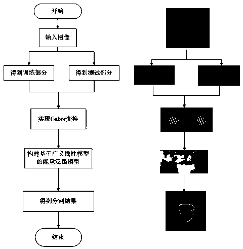
[0037] The medical image lesion region segmentation method based on the energy functional model of machine learning of the present invention is as follows: image 3 As shown in the middle left half, follow the steps below:

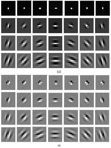
[0038] Step 1: Initialize the parameter value of the feature array of the Gabor transform, and set the feature array to zero; at the same time, take the upper 1 / 2 part of the input medical image as the test set, and the lower 1 / 2 part as the training set, respectively for the training set The training image and the test image in the test set are subjected to Gabor transformation, and the general expression of the Gabor transformation is as follows:

[0039] plural form:

[0040]

[0041] Real part:

[0042]

[0043] Imaginary part:

[0044]

[0045] In formulas (1), (2), and (3), λ represents the wavelength of the sine wave, θ represents the direction of the filter, ψ represents the initial phase, σ represents the standard deviation of the Gaus...


PUM


Abstract

The invention discloses a medical image lesion area segmentation method based on an energy functional model of machine learning, and the method comprises the steps: firstly processing a medical imagethrough employing Gabor transformation and a generalized linear machine learning method, capturing the discrimination description of a lesion area and a normal tissue area, and obtaining a learning model based on the lesion area; and then constructing the energy functional model on the basis, and segmenting the lesion area of the medical image. The problems that a machine learning method is sensitive to label data and the energy functional can fall into a local minimum value are solved, the medical image with a complex background can be well segmented, and the feature information of the lesionarea is effectively reserved and reliable guarantee is provided for accuracy of diagnosis of the lesion area for the doctor.

Description

technical field [0001] The invention relates to the field of medical image segmentation, in particular to a medical image lesion area segmentation method based on an energy functional model of machine learning. Background technique [0002] In 2011, the American medical community first proposed the "precision medicine" program, which combines human cognition of disease mechanisms and related medical data information technology to accurately classify and diagnose diseases. , more precise medical methods. However, due to factors such as differences in medical imaging equipment and complex internal structures of the human body, characteristics such as uneven gray distribution, blurred edges, and high noise intensity usually appear when acquiring medical images. The existing automatic segmentation methods for medical images are not capable of resolving the tissue of the lesion area enough, and thus cannot accurately predict the boundary of the lesion area in the image. In addi...

Claims


Application Information

Patent Timeline
Patent Type & Authority Applications(China)
IPC IPC(8): G06T7/00G06T7/11
CPCG06T7/0012G06T7/11G06T2207/20081G06T2207/20084G06T2207/30096Y02T10/40
Inventor 方玲玲赵欣怡
Owner LIAONING NORMAL UNIVERSITY
Why Eureka
Try Eureka