
Ultrasound imaging equipment, system and image enhancement method for contrast-enhanced ultrasound imaging

A technology of image enhancement and contrast-enhanced ultrasound, which is applied in image enhancement, ultrasound/acoustic/infrasonic diagnosis, image analysis, etc. It can solve the problems of decreased lateral resolution of imaging, misdiagnosis by doctors, and inability of doctors to judge strong signals, etc.

Active Publication Date: 2022-07-22
BEIJING SHEN MINDRAY MEDICAL ELECTRONICS TECH RES INST +2
View PDF18 Cites 0 Cited by
  • Summary
  • Abstract
  • Description
  • Claims
  • Application Information

AI Technical Summary

Problems solved by technology

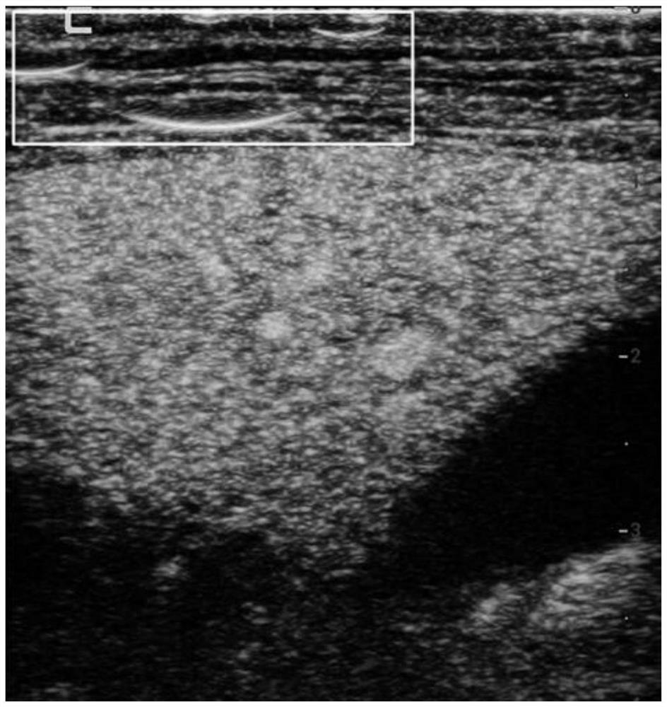
However, arc artifacts caused by signal saturation will appear in plane wave contrast images, such as figure 1 shown in the white box
In the process of ultrasonic diagnosis, the arc artifact will bring trouble to the doctor, making it difficult for the doctor to judge whether the strong signal is the ultrasonic echo reflected by the biological tissue under test or interference, which may lead to misdiagnosis by the doctor
[0004] In order to eliminate arc artifacts, on the one hand, the receiving aperture can be reduced or the receiving apodization coefficient can be adjusted to control it, but this solution will broaden the main lobe of the signal, resulting in a decrease in the lateral resolution of imaging; on the other hand, it can reduce Analog gain to minimize saturation at the expense of signal strength

Method used


Image

  • Ultrasound imaging equipment, system and image enhancement method for contrast-enhanced ultrasound imaging
  • Ultrasound imaging equipment, system and image enhancement method for contrast-enhanced ultrasound imaging
  • Ultrasound imaging equipment, system and image enhancement method for contrast-enhanced ultrasound imaging

Examples


Embodiment Construction

[0053] The present invention will be further described in detail below through specific embodiments in conjunction with the accompanying drawings. Wherein similar elements in different embodiments have used associated similar element numbers. In the following embodiments, many details are described so that the present application can be better understood. However, those skilled in the art will readily recognize that some of the features may be omitted in different circumstances, or may be replaced by other elements, materials, and methods. In some cases, some operations related to the present application are not shown or described in the specification, in order to avoid the core part of the present application being overwhelmed by excessive descriptions, and for those skilled in the art, these are described in detail. The relevant operations are not necessary, and they can fully understand the relevant operations according to the descriptions in the specification and general ...


PUM


Abstract

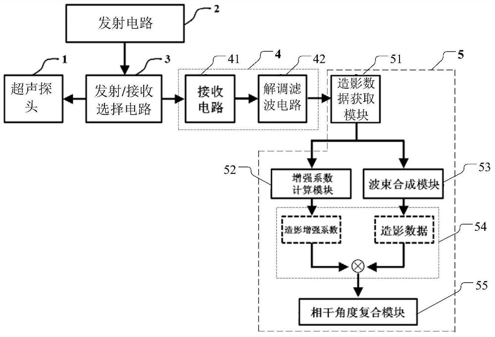
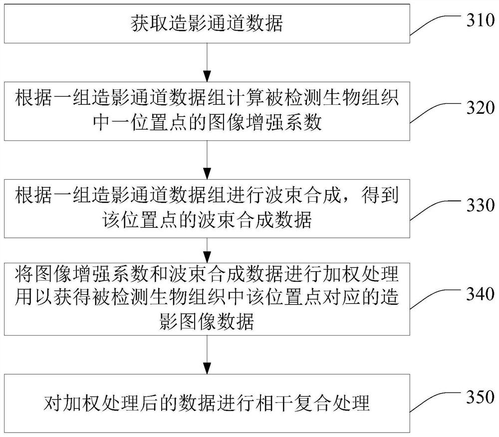
An ultrasound imaging device, a system and an image enhancement method for contrast-enhanced ultrasound imaging thereof. Because the image enhancement coefficients of each position point in the detected biological tissue are first calculated according to the contrast channel data, and then the image enhancement coefficients and the beamforming data are weighted.

Description

technical field [0001] The invention relates to an ultrasound imaging device, in particular to an image enhancement technology for contrast-enhanced ultrasound imaging in the ultrasound imaging device. Background technique [0002] Existing contrast-enhanced ultrasound imaging uses the traditional focused emission mode. Taking the most basic single beam as an example, only one receiving line can be formed for each transmission; even if multi-beam parallel processing is used, the number of transmissions cannot be significantly reduced. For contrast-enhanced ultrasound imaging, more firing times means that the excitation intensity of the microbubbles in a unit frame by the sound field increases, which will inevitably lead to a shortening of the duration of the contrast-enhanced image, thereby affecting the integrity of blood perfusion at the lesion. Although the purpose of prolonging the duration can be achieved by reducing the number of transmissions or reducing the intensit...

Claims


Application Information

Patent Timeline
Patent Type & Authority Patents(China)
IPC IPC(8): A61B8/00
CPCA61B8/06A61B8/481A61B8/5207G06T5/50G06T7/0012G06T2207/10132G06T2207/20056G06T5/90
Inventor 章希睿唐杰罗渝昆张明博桑茂栋向兰茜覃东海范伟
Owner BEIJING SHEN MINDRAY MEDICAL ELECTRONICS TECH RES INST
Learn More