Three-dimensional ultrasonic diagnosis device for gynecological diseases

A diagnostic device and technology for gynecological diseases, applied in the fields of sonic diagnosis, infrasonic diagnosis, ultrasonic/sonic/infrasonic diagnosis, etc., which can solve problems such as difficulty in establishing 3D models of 2D ultrasonic images, difficulty in gynecological disease diagnosis, poor consistency, etc. Achieve the effect of convenient and efficient detection process, accurate and efficient 3D reconstruction, and comprehensive detection methods

Inactive Publication Date: 2020-07-24
王时灿
View PDF18 Cites 2 Cited by
  • Summary
  • Abstract
  • Description
  • Claims
  • Application Information

AI Technical Summary

Problems solved by technology

However, at present, the acquisition of clinical ultrasound image information is mainly obtained by the doctor's hand-held ultrasound probe, which has problems of unstable operation and poor consistency. It is difficult to accurately acquire two-dimensional ultrasound images of each section, resulting in two-dimensional Ultrasound imaging is difficult and inaccurate when building a three-dimensional model, which affects the accuracy of the final diagnostic result
[0005] In addition, because there are many types of gynecological diseases, some diseases not only do not show obvious specificity in clinical symptoms, but also have relatively similar ultrasound images, which makes it difficult to diagnose gynecological diseases based on such ultrasound images, and it is difficult to accurately identify them. Types of diseases, resulting in inaccurate and comprehensive diagnostic results

Method used


Image

  • Three-dimensional ultrasonic diagnosis device for gynecological diseases
  • Three-dimensional ultrasonic diagnosis device for gynecological diseases
  • Three-dimensional ultrasonic diagnosis device for gynecological diseases

Examples


Embodiment

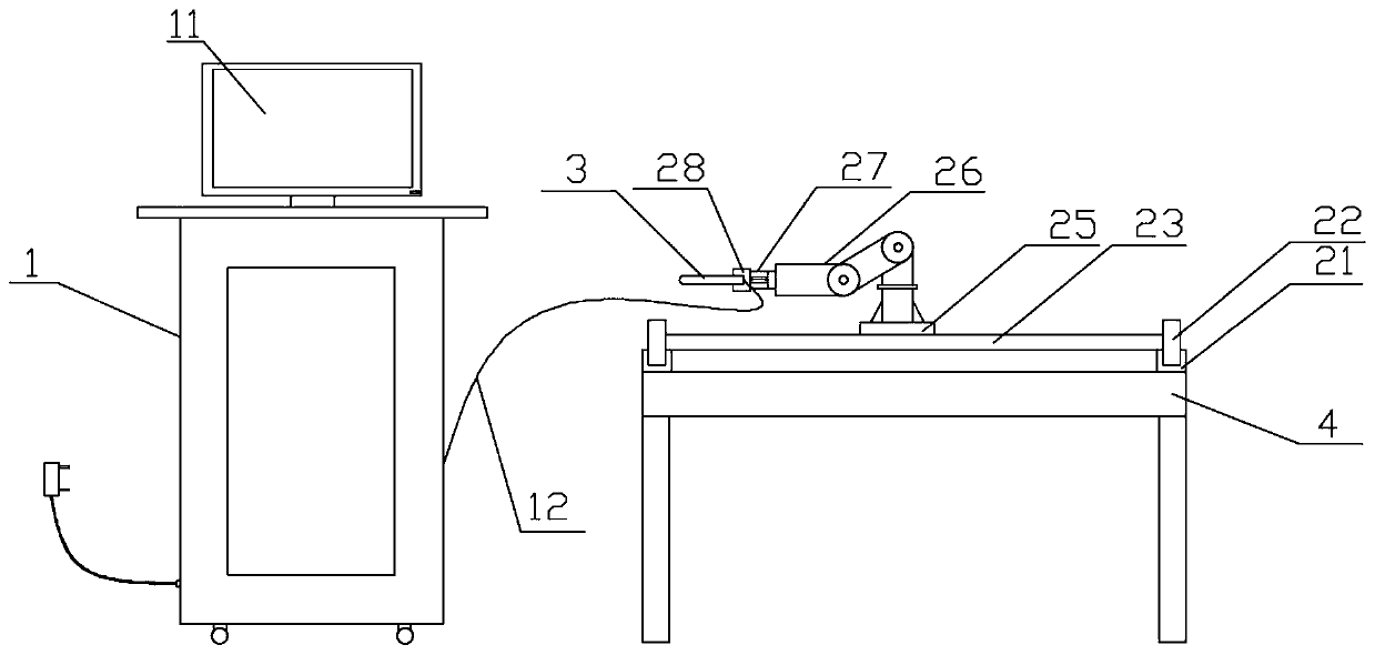
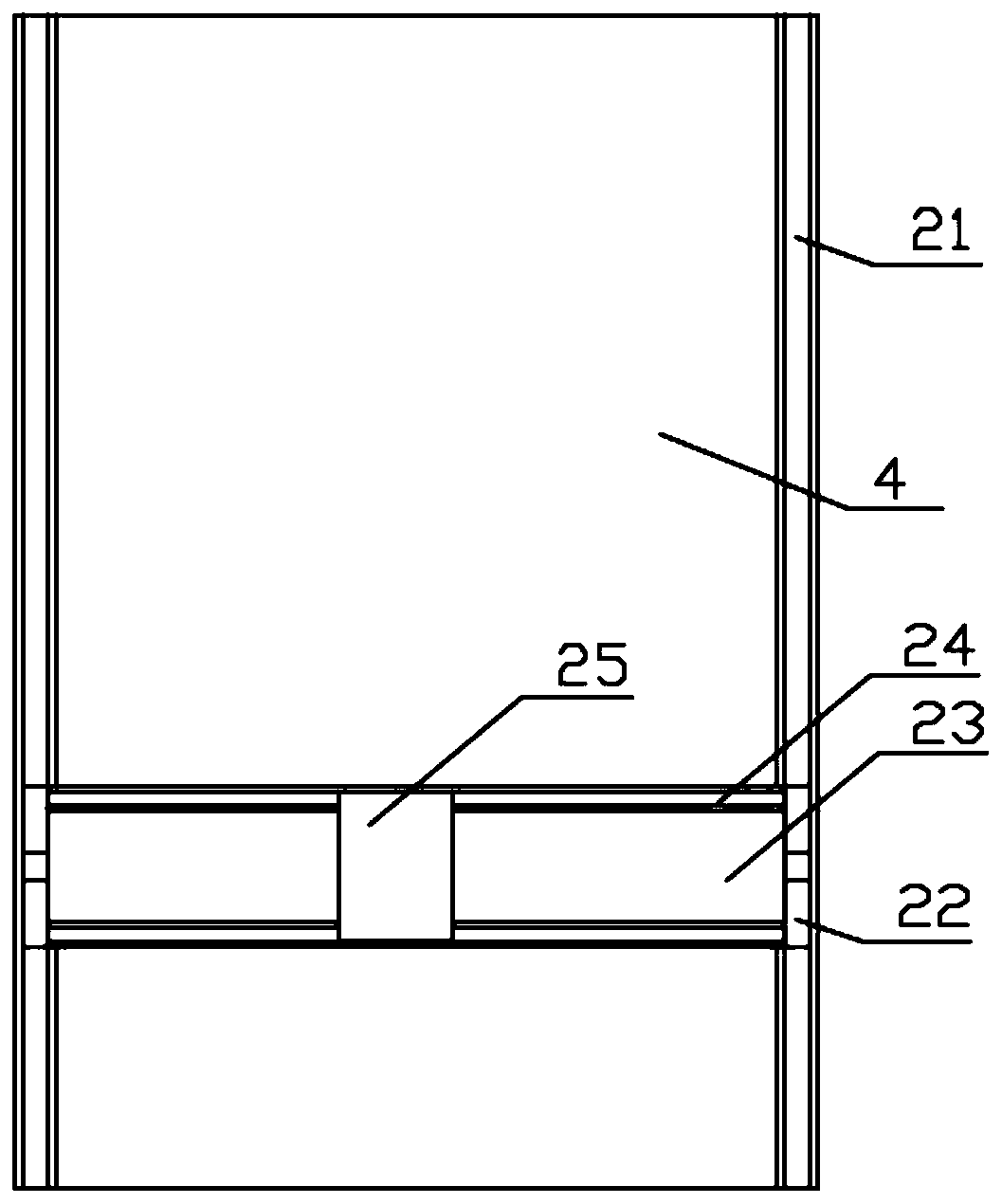
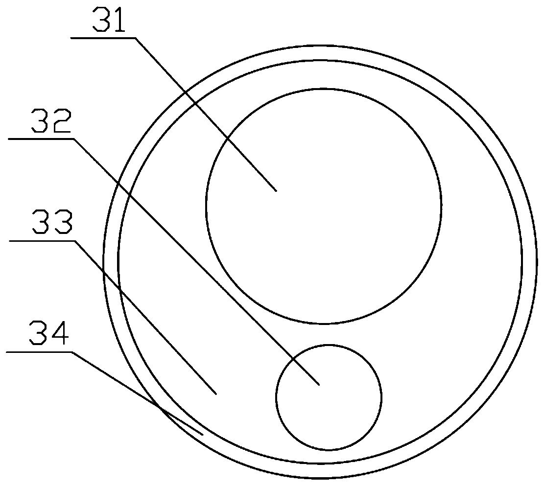
[0030] see figure 1 , the embodiment of the present invention provides a three-dimensional ultrasonic diagnosis device for gynecological diseases, including a diagnostic device host 1, a mechanical scanning mechanism and a detection probe 3, and the mechanical scanning mechanism establishes communication with the diagnostic device host 1 based on the TCP / IP communication protocol connected, the detection probe is electrically connected to the host of the diagnostic device through a connection line 12; the mechanical scanning mechanism is fixed on the detection bed 4, and the detection probe 3 is detachably installed on the mechanical scanning mechanism for Automatic scanning is carried out under the drive of the mechanical scanning mechanism; an ultrasonic probe 31 and an optical lens 32 are arranged in the detection probe 3, which are respectively used to collect two-dimensional ultrasonic images and optical images of the patient; A gynecological disease diagnosis system is p...


PUM


Abstract

The invention discloses a three-dimensional ultrasonic diagnosis device for gynecological diseases. The device comprises a diagnosis device main machine, a mechanical scanning mechanism and a detection probe, and the detection probe is driven by the mechanical scanning mechanism to perform automatic scanning; an ultrasonic probe and an optical lens are arranged in the detection probe and are respectively used for acquiring a two-dimensional ultrasonic image and an optical image of a patient; and a gynecological disease diagnosis system is arranged in the diagnosis device main machine and is used for controlling the mechanical scanning mechanism and processing and analyzing the acquired ultrasonic images and optical images. By means of the mode, the detection probe can be accurately controlled to conduct intracavity ultrasonic diagnosis, so that the obtained two-dimensional image has position information, and accurate three-dimensional reconstruction is facilitated; and in addition, theoptical image and the ultrasonic image can be fused, the resolution of the ultrasonic image is effectively improved, so that a finer tissue structure is obtained, a lesion area is segmented and extracted more accurately, and accurate diagnosis of the gynecological diseases is achieved.

Description

technical field [0001] The invention relates to the technical field of ultrasonic diagnosis, in particular to a three-dimensional ultrasonic diagnosis device for gynecological diseases. Background technique [0002] Gynecological diseases, as common and frequently-occurring diseases of women, need timely and effective treatment to avoid the impact of prolongation or deterioration of the course of disease on patients. However, because many diseases in gynecological diseases do not have specific symptoms, effective and accurate diagnosis in time is of great significance for the discovery and treatment of diseases. Among various clinical diagnostic methods for gynecological diseases, intracavitary ultrasound diagnosis has been widely used as a non-invasive, painless, convenient, intuitive and effective inspection method. [0003] At present, two-dimensional ultrasound is often used in clinical diagnosis of gynecological diseases to effectively obtain images of lesion locations...

Claims


Application Information

Patent Timeline
IPC IPC(8): A61B8/12A61B8/08A61B8/00A61B1/303A61B1/04A61B1/07A61B1/06A61B1/00
CPCA61B1/00009A61B1/00149A61B1/04A61B1/0661A61B1/07A61B1/303A61B8/08A61B8/12A61B8/4218A61B8/483A61B8/5261
Inventor 王时灿
Owner 王时灿
Why Eureka
Try Eureka