Short leading and trailing frames to improve image quality in positron emission tomography (PET)

A leading frame and electronic technology, applied in radiation measurement, 2D image generation, computer tomography scanner, etc., can solve the problems of inaccurate modeling of active scattering, useful AFOV reduction, and large edge slice noise. Achieve improved sensitivity, reduced scan time, and better coverage

Pending Publication Date: 2020-07-31
KONINKLJIJKE PHILIPS NV
View PDF0 Cites 1 Cited by
  • Summary
  • Abstract
  • Description
  • Claims
  • Application Information

AI Technical Summary

Problems solved by technology

[0003] However, in the case of existing hybrid PET / computed tomography (PET / CT) scanners with an AFOV coverage of only 15 cm, edge slices appear much noisier than central slices due to reduced geometric sensitivity of the edge regions. many
Thus the effective useful AFOV is further reduced by
Also, due to the limited AFOV, it is not possible to accurately model scatter from activity in target parts of the patient, such as the liver

Method used


Image

  • Short leading and trailing frames to improve image quality in positron emission tomography (PET)
  • Short leading and trailing frames to improve image quality in positron emission tomography (PET)
  • Short leading and trailing frames to improve image quality in positron emission tomography (PET)

Examples


Embodiment Construction

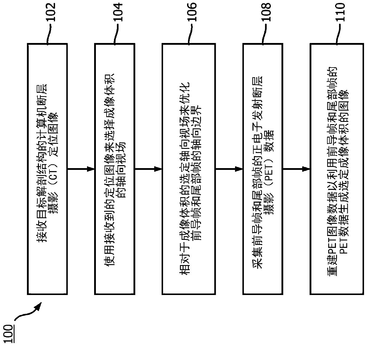
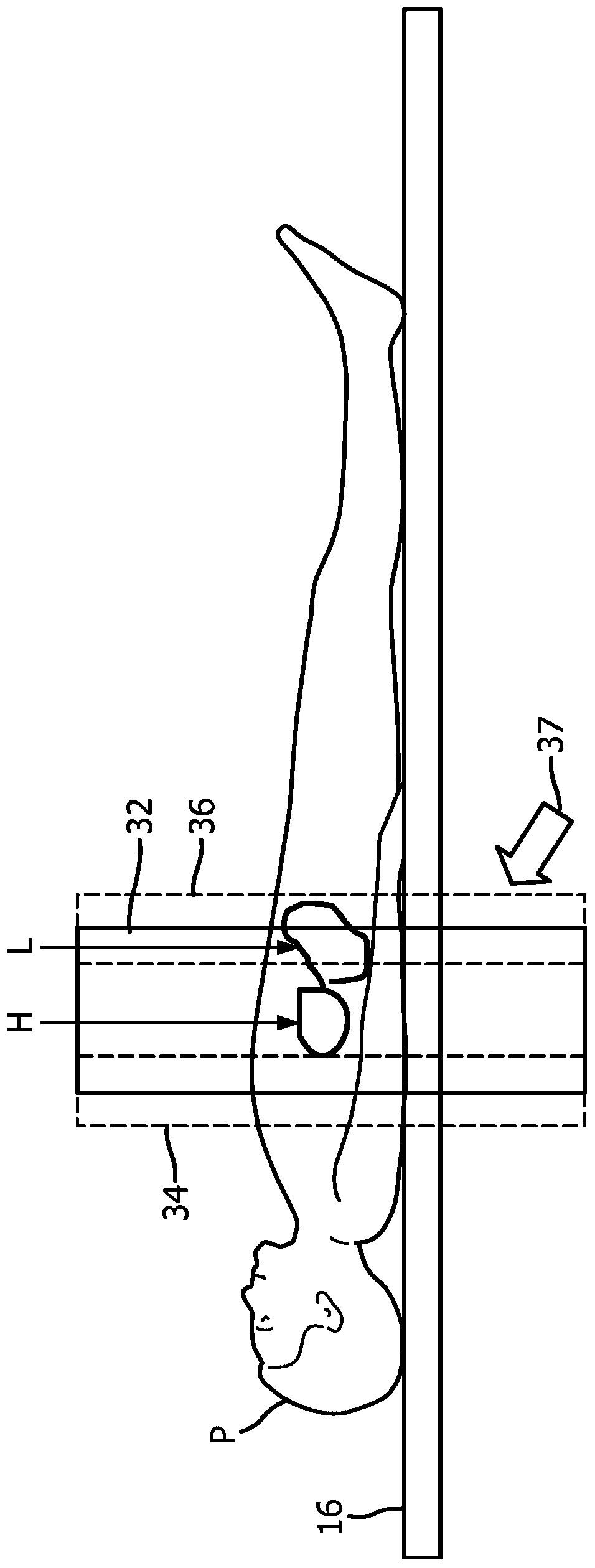
[0022] The present disclosure relates to improvements in PET imaging using one or more axial frames. In an illustrative contemplated embodiment, cardiac PET imaging is performed using PET / CT with a single frame having a system axial extent of about 15 cm. In practice, it was found that edge effects at the axial ends of the FOV could cause the outer edge axial slices to be degraded or unusable, resulting in an effective axial width of approximately 12 cm. This is barely large enough to accommodate a normal heart, which may truncate the heart if it is larger than normal or if the axial positioning of the heart in the PET scanner is less than fully centered in the FOV in the axial direction. The degradation of edge slices has two causes: (1) scattering from outside the FOV, and (2) low sensitivity (where the sensitivity of edge slices can be seen as the difference between the total count of edge slices and the axial slice located at the center of FOV Ratio compared to the total ...


PUM


Abstract

A non-transitory computer-readable medium storing instructions readable and executable by a workstation (18) including at least one electronic processor (20) to perform an image reconstruction method(100). The method includes: receiving a surview image of a target anatomy acquired by an image acquisition device (12); selecting an axial field of view (FOV) of an imaging volume using the received surview image; optimizing axial boundaries of leading and trailing frames respective to the selected axial FOV of the imaging volume; operating a positron emission tomography (PET) imaging device (14)to acquire PET imaging data including acquiring PET imaging data for the leading frame followed by acquiring PET imaging data for the imaging volume followed by acquiring PET imaging data for the trailing frame; and reconstructing the PET imaging data to generate an image of the selected imaging volume including out-of-axial FOV scatter correction performed using the PET imaging data acquired forat least one of the leading and trailing frames.

Description

technical field [0001] The following generally relates to medical imaging techniques, medical image interpretation techniques, image reconstruction techniques, and related techniques. Background technique [0002] For cost-saving reasons, positron emission tomography (PET) scanners typically have a relatively small axial field of view (AFOV). When the PET scan covers only the portion of the patient's body between the head and the feet, the acquired data is essentially "truncated" in the axial direction, resulting in an image at and near the first and last axial slices The quality and quantification of the damage. For example, a heart scan can be done using a single frame. [0003] However, in the case of existing hybrid PET / computed tomography (PET / CT) scanners with an AFOV coverage of only 15 cm, edge slices appear much noisier than central slices due to reduced geometric sensitivity of the edge regions. many. The effective useful AFOV is thus further reduced. Also, du...

Claims


Application Information

Patent Timeline
Patent Type & Authority Applications(China)
IPC IPC(8): A61B6/03A61B6/00G01T1/29G06T11/00
CPCA61B6/037A61B6/4417A61B6/488A61B6/503A61B6/5205A61B6/5282A61B6/545G06T11/005G01T1/2985
Inventor A·安德烈耶夫白传勇宋犀云叶京汉
Owner KONINKLJIJKE PHILIPS NV
Why Eureka
Try Eureka