Visual electronic photoacoustic endoscope for urethral stenosis

A urethral stricture, visual electronic technology, applied in the direction of endoscope, urethroscope, cystoscope, etc., can solve the problems of inaccurate urethral dilator expansion amount, inability to accurately judge the severity of urethral stricture, etc., and achieve accurate urethral dilator. Dilation volume, the effect of overcoming the dilation volume of the urethral dilator

Pending Publication Date: 2020-08-07
陈征
View PDF0 Cites 0 Cited by
  • Summary
  • Abstract
  • Description
  • Claims
  • Application Information

AI Technical Summary

Problems solved by technology

[0006] The purpose of the present invention is to solve the above-mentioned problems in the prior art, and the present invention provides a visual electronic photoacoustic endoscopic device for urethral stricture, which includes an ultrasonic detection module, an operation conversion module, an imaging module, a matching module and an implant catheter. The ultrasonic detection module collects ultrasonic signals, the ultrasonic signals are generated by the urethral stricture irradiated by the pulsed laser, the operation conversion module converts the ultrasonic signals ...

Method used


Image

  • Visual electronic photoacoustic endoscope for urethral stenosis
  • Visual electronic photoacoustic endoscope for urethral stenosis
  • Visual electronic photoacoustic endoscope for urethral stenosis

Examples


example 1

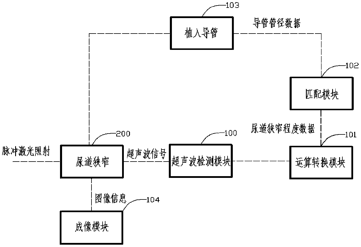
[0041] Such as figure 1As shown, the visualized endoscopic device of this embodiment includes: an ultrasonic detection module 100, an operation conversion module 101, an imaging module 104, a matching module 102, and an implant catheter 103. The ultrasonic detection module 100 collects ultrasonic signals, and the ultrasonic signals Produced by the urethral stricture 200 irradiated by the pulsed laser, the operation conversion module 101 converts the ultrasonic signal into the data of the degree of urethral stricture, the imaging module 104 displays the image of the urethral stricture, and the implanted catheter 103 matches The module 102 outputs the implanted catheter diameter data according to the urethral stricture degree data. The urethral stricture is irradiated by pulsed laser to generate ultrasonic signals based on the following principle: Photoacoustic Imaging (PAI) is a new non-invasive and non-ionizing biomedical imaging method developed in recent years. When the pul...

Embodiment 3

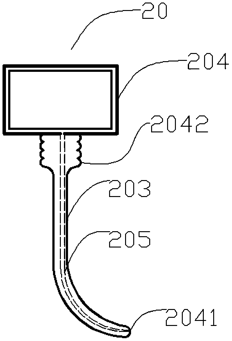
[0043] figure 2 A schematic diagram of a visualized endoscopic product 20 according to this embodiment is shown (the ultrasonic detection module 100 , the operation conversion module 101 and the matching module 102 are not shown). Wherein, the imaging module includes: a display device 204 , an implant catheter 203 , an optical signal channel 205 , a light source 2041 , a mirror (not shown), and a handle 2042 . Wherein, the outside of the optical signal channel 205 is covered with an expandable device. Segmental regulation of implant catheter 203 can be achieved through compression and / or expansion of handle 2042 .

[0044] Implementation 3:

[0045] The difference compared with Embodiment 1 is that the image data received by the imaging module is an image obtained after restoration of the degree of urethral stricture data converted by the operation conversion module 101, and / or the matching module includes an inflation control module , the implantation catheter is a bendab...

Embodiment 4

[0047] Such as Figure 4 Said, the present embodiment also has a signal amplifying circuit module 105 and / or data storage module (not shown) on the basis of embodiment 1, and the signal amplifying circuit module 105 carries out the ultrasonic signal detected by the ultrasonic detecting module. After denoising and amplification processing, it is output to the operation conversion module 101 . The signal amplifying circuit module 105 may be a commonly used signal amplifying circuit in the field of ultrasonic signal amplifying, which will not be repeated here. The operation conversion module 101 converts the ultrasonic signal into the urethral stricture degree data and stores it in the data storage module.


PUM


Abstract

The invention provides a visual electronic photoacoustic endoscopic device. The device comprises a visual screen, an electronic mirror, a signal external connection device, an operation output unit, amathematical calculation conversion system, an amplification circuit, an imaging system and an implantable expandable air bag. The visual electronic photoacoustic endoscopic device mainly solves theproblems that in the prior art, the severity and depth of urethral stenosis cannot be accurately judged, and the dilation amount of the urethral dilator cannot be accurately determined, and achieves the technical effects of quantitatively observing the severity and depth of urethral stenosis and accurately determining the dilation amount of the urethral dilator.

Description

technical field [0001] The invention is used in the technical field of medical instruments, and in particular relates to a visual electronic photoacoustic endoscope for urethral stricture. Background technique [0002] With the continuous progress of minimally invasive treatment, trauma and iatrogenic urethral injury have become the main factors of urethral stricture. [0003] Known methods for diagnosing urethral stricture include: urethrography, urethral cystoscopy, urethroscopy, ultrasonography, etc., but none of them can accurately determine the severity and depth of urethral stricture. [0004] In addition, the existing urethral dilation devices are usually blind dilation, which cannot accurately dilate the amount according to the degree of urethral stenosis, resulting in frequent blood flow during the operation, which brings great trauma to the patient's body and mind. [0005] The inventor has not found a technical solution that can better solve the above problems. ...

Claims


Application Information

Patent Timeline
IPC IPC(8): A61B1/307A61B1/04A61B5/00A61B8/12
CPCA61B1/04A61B1/307A61B5/0095A61B5/6852A61B5/6867A61B5/7225A61B8/12
Inventor 陈征王珏
Owner 陈征
Why Eureka
Try Eureka