
Measurement guidance for coronary flow estimation from bernoulli's principle
An endometry, coronary technique, applied in the field of computer programs, which can solve the problems of lack of robustness, increased complexity, etc.
Patent Information
- Authority / Receiving Office
- CN · China
- Patent Type
- Applications(China)
- Current Assignee / Owner
- Publication Date
- 2020-09-11
Smart Images
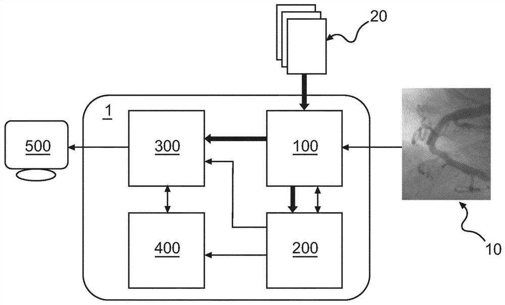
Figure 1 
Figure 2 
Figure 3
Abstract
Description
technical field
[0001] The invention relates to a device for assessing hemodynamic properties in the coronary vasculature, a corresponding method and a corresponding computer program. In particular, the present invention relates to improved measurement guidance for performing measurements of hemodynamic parameters by deriving from diagnostic data of the vasculature optimal measurement positions for intravascular measurements. Background technique
[0002] The severity of functional stenosis in coronary arteries can usually be assessed by means of intravascular pressure measurements. In this regard, one can in particular consider the pressure at the distal end of the stenosis (P d ) and the pressure in the aorta (P a ) to perform the evaluation. Thus, this ratio can be taken into account using metrics such as Fractional Flow Reserve (FFR) or Instantaneous Wave-Free Ratio (iFR), both of which are determined from said pressure ratio. FFR measurements use pressure curves thr...
Examples
Embodiment Construction
[0115] The illustrations in the figures are schematic. In different figures, similar or identical elements bear the same reference signs.
[0116] figure 1 An exemplary embodiment of a device 1 for assessing hemodynamic properties is schematically represented. The device 1 includes an input unit 100 , a recognition unit 200 , a calculation unit 300 , a modeling unit 400 and a display unit 500 .
[0117] The input unit 100 receives a diagnostic image 10 of a vessel of interest. in accordance with figure 1 In the exemplary embodiment of the present invention, the diagnostic image 10 is a two-dimensional image acquired using X-ray angiography. The input unit 100 provides the diagnostic image 10 to the recognition unit 200 .
[0118] The recognition unit 200 is configured to receive the diagnostic image 10 and perform vessel segmentation of the vessel of interest represented in the diagnostic image 10, i.e. the recognition unit 200 segments the (image of) the vessel of intere...