Head and neck lymph node and drainage area automatic sketching method based on deep learning

A lymphatic drainage and deep learning technology, applied in the field of medical images, can solve problems such as inability to support segmentation model training, delineation dependencies, and blood vessel confusion.

Active Publication Date: 2020-10-30
PERCEPTION VISION MEDICAL TECH CO LTD
View PDF5 Cites 7 Cited by
  • Summary
  • Abstract
  • Description
  • Claims
  • Application Information

AI Technical Summary

Problems solved by technology

The lymphatic drainage area has good contrast and boundaries on CT, but lymph nodes are difficult to distinguish on localized CT or enhanced CT for radiotherapy, especially easily confused with adjacent muscles and blood vessels, resulting in mis-outlined or missed outlines
[0003] At present, the boundaries of metastatic lymph nodes and lymphatic drainage areas are mainly drawn manually by doctors or image segmentation methods based on deep learning, in which the efficiency of manual drawing is low, the repeatability is poor, and it is heavily dependent on the experience level of drawing doctors and drawing standard references
In addition, for different types of tumors and tumors in different locations in the head and neck, the risk of lymph node metastasis is quite different, which brings greater challenges to the clinical delineation of metastatic lymph nodes.
[0004] However, the existing conventional image segmentation methods based on deep learning rely on image labeling and training with a large amount of data, and clinically obtained data usually have problems with incomplete labeling and inconsistent labeling schemes, resulting in the actual amount of training data available. rarely
Therefore, the existing deep learning-based technology is still unable to support the automatic delineation of low-contrast and irregularly distributed lymph nodes.
In addition, the lymphatic drainage area of ​​the head and neck covers a large range of images, and its delineation depends on the image information between layers. The general hardware configuration cannot support the training of segmentation models under large-scale 3D image input.

Method used


Image

  • Head and neck lymph node and drainage area automatic sketching method based on deep learning

Examples


Embodiment Construction

[0034] In order to make the purposes, technical solutions and advantages of the embodiments of the present invention clearer, the technical solutions in the embodiments of the present invention will be clearly and completely described below with reference to the accompanying drawings in the embodiments of the present invention. Obviously, the described embodiments These are some embodiments of the present invention, but not all embodiments. Based on the embodiments of the present invention, all other embodiments obtained by those of ordinary skill in the art without creative efforts shall fall within the protection scope of the present invention.

[0035] Unless expressly stated otherwise, throughout the specification and claims, the term "comprising" or its conjugations such as "comprising" or "comprising" and the like will be understood to include the stated elements or components, and Other elements or other components are not excluded.

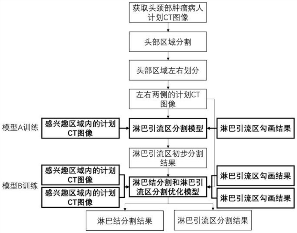
[0036] figure 1 A flow chart of a...


PUM


Abstract

The embodiment of the invention provides a head and neck lymph node and drainage area automatic sketching method based on deep learning. The method utilizes the symmetry of a human body structure to divide a head area into a left part and a right part for training and prediction, thereby indirectly increasing the data volume of model training, and reducing the scale of a deep learning model at thesame time. According to the method, a step-by-step processing optimized sketching strategy is used, the lymphatic drainage area easy to segment is achieved through the deep learning model, then the lymphatic drainage area is optimized and lymph nodes are segmented through the multi-task deep learning model, the relevance between the lymphatic drainage area and the lymph nodes is fully utilized, and the segmentation accuracy of the lymphatic drainage area and the lymph nodes is improved. According to the invention, an AI (artificial intelligence)-assisted contour sketching method is implemented in a radiotherapy plan work flow, so that the sketching consistency of the work efficiency of medical workers can be effectively improved, and the head and neck tumor radiotherapy precision is improved.

Description

technical field [0001] The invention relates to the field of medical images, in particular to an automatic delineation method for head and neck lymph nodes and drainage areas based on deep learning. Background technique [0002] Due to the extensive distribution of lymphatic network in the head and neck mucosa, the risk of regional lymph node metastasis of head and neck tumors is high, and the probability of occult metastasis is as high as 30%. In the clinical radiotherapy of patients with head and neck tumors, in addition to high-dose irradiation to the primary tumor (GTV), preventive irradiation is also required to the lymph nodes with metastases (GTVn) and lymphatic drainage areas (CTVn) around the tumor . At present, radiotherapy is one of the important treatment methods for head and neck tumors, and there are many important and delicate structures in the head and neck. Accurate radiotherapy technology has important clinical significance for the quality of life of patie...

Claims


Application Information

Patent Timeline
IPC IPC(8): G06T7/00G06T7/11G16H30/40
CPCG06T7/0012G06T7/11G16H30/40G06T2207/10081G06T2207/20081G06T2207/30004G06T2207/20104
Inventor 孙颖陆遥林丽陈海斌
Owner PERCEPTION VISION MEDICAL TECH CO LTD
Why Eureka
Try Eureka