Retinal vessel segmentation algorithm based on level set

A retinal blood vessel and segmentation algorithm technology, applied in the field of medical image segmentation, can solve the problems of difficult detection of tiny blood vessels and weak anti-noise ability

Pending Publication Date: 2020-11-06
HARBIN UNIV OF SCI & TECH
View PDF0 Cites 2 Cited by
  • Summary
  • Abstract
  • Description
  • Claims
  • Application Information

AI Technical Summary

Problems solved by technology

[0005] The purpose of the present invention is to provide a retinal blood vessel segmentation algorithm based on level sets, through which the problems of difficult detection of small blood v

Method used


Image

  • Retinal vessel segmentation algorithm based on level set
  • Retinal vessel segmentation algorithm based on level set
  • Retinal vessel segmentation algorithm based on level set

Examples


Embodiment 1

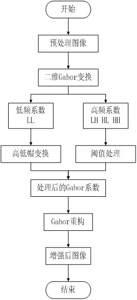
[0041] A level set based retinal vessel segmentation algorithm, the algorithm comprises the following steps:

[0042]Step 1. Retinal image preprocessing: including single-channel color acquisition, region-of-interest extraction, and brightness adjustment. In a color retinal image, compared with other color channels, blood vessels have the best contrast between their green channel and the background area , the green channel in the fundus color retinal image is used as the input image for subsequent blood vessel segmentation. Mask extraction is to cover the processed image with the selected image and control the image processing area. Due to the gray value of the edge pixel and the gray value of the ROI pixel There is a large difference between the degree values. When extracting the mask, the threshold binarization method is used. The brightness enhancement adopts the contrast-limited adaptive histogram equalization, divides the image into blocks, and calculates the histogram in ...


PUM


Abstract

The invention relates to a retinal vessel segmentation algorithm based on a level set. At present, in medical clinic, fundus images are an important basis for ophthalmologists to diagnose and treat patients with fundus diseases, and the doctors screen the fundus images and judge lesion types by means of subjective experience so as to give diagnosis. This said medical mode consumes a lot of time, has subjectivity and is easy to miss the optimal treatment time. The method of the invention comprises the following steps: preprocessing a retina image, including single-channel color acquisition, region-of-interest extraction and brightness adjustment; performing retinal image enhancement: enhancing a retinal blood vessel image by adopting a method based on combination of Gabor transformation andhigh-low cap transformation; performing retinal vessel segmentation: using an active contour model mainly, using smooth and closed contour lines for covering the edges of sub-pixels, and obtaining anaccurate segmentation target through energy functional minimization of the active contour model. The invention is used for the retinal vessel segmentation algorithm based on the level set.

Description

Technical field: [0001] The invention relates to the technical field of medical image segmentation, in particular to a retinal blood vessel segmentation algorithm based on a level set. Background technique: [0002] In recent years, with the rapid development of image processing and analysis technology, medical image processing by computer has been widely used in various disciplines and fields of medicine. With the help of image processing, computer vision, and machine learning technologies, the processing and analysis of relevant medical images can effectively quantify and visualize relevant pathological and anatomical structures, so as to realize computer-assisted or even replace doctors for accurate diagnosis and precise treatment of diseases. [0003] The visual system is an important tool for people to obtain external information and plays an irreplaceable role in work, study and life. The health of the eyes is inseparable from people's quality of life. According to st...

Claims


Application Information

Patent Timeline
IPC IPC(8): G06T7/11G06T7/13G06T7/136G06T7/90G06T5/00G06T5/40
CPCG06T5/002G06T5/40G06T2207/10024G06T2207/30041G06T2207/30101G06T7/11G06T7/13G06T7/136G06T7/90
Inventor 王英立侯晓晓
Owner HARBIN UNIV OF SCI & TECH
Why Eureka
Try Eureka