Protein chip for rapidly detecting pleuroperitoneal carcinoma cells and preparation method thereof

A protein chip and cancer cell technology, applied in the field of biomedicine, can solve the problems of lower detection accuracy, insufficient detection accuracy, and inability to use antibodies, etc., and achieve the effect of improving detection accuracy

Pending Publication Date: 2020-11-27
深圳市森盈生物科技有限公司
View PDF5 Cites 0 Cited by
  • Summary
  • Abstract
  • Description
  • Claims
  • Application Information

AI Technical Summary

Problems solved by technology

However, in the prior art, since cell smears are not as reproducible as tissue sections, the cell components and distribution on each cell smear are different, so there is no guarantee that there are cancer cells on each smear , so that

Method used


Image

  • Protein chip for rapidly detecting pleuroperitoneal carcinoma cells and preparation method thereof

Examples


Embodiment Construction

[0020] Below in conjunction with accompanying drawing and embodiment the present invention will be further described:

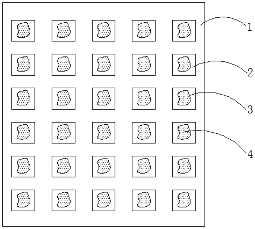
[0021] refer to figure 1 : A protein chip for rapid detection of pleural effusion cancer cells, comprising a glass slide 1, the glass slide 1 is an aldylated glass slide, and the slide glass 1 is divided into a plurality of microarray areas 2 by means of a square array , each microarray region 2 is attached with carboxyl magnetic beads 3, and the surface of carboxyl magnetic beads 3 is irregular, so it has a relatively large surface area, which can increase the contact probability between carboxyl magnetic beads 3 and coupling molecules, and improve coupling efficiency , colloidal gold-labeled anti-tumor antigen-specific antibody 4 is cross-linked inside the carboxyl magnetic beads 3, and the anti-tumor antigen-specific antibody 4 is carcinoembryonic antigen, carbohydrate antigen, carbohydrate antigen, epithelial-specific antigen and epithelial membrane antig...


PUM


Abstract

The invention provides a protein chip for rapid detection of pleuroperitoneal carcinoma cells and a preparation method of the protein chip, and relates to the technical field of biomedicine. Accordingto the protein chip for rapidly detecting the pleuroperitoneal carcinoma cells and the preparation method of the protein chip, the protein chip comprises a glass slide, the glass slide is divided into a plurality of micro-array areas in a square array mode, and carboxyl magnetic beads are attached to the interior of each micro-array area. According to the protein chip for rapid detection of pleuroperitoneal carcinoma cells and a preparation method of the protein chip, carboxyl magnetic beads are attached to a glass slide; meanwhile, the carboxyl magnetic beads and the colloidal gold labeled antibody are subjected to crosslinking; the carboxyl magnetic beads and the colloidal gold labeled antibodies are subjected to cross-linking, and the sample application is performed in each micro-arrayregion, so that the colloidal gold labeled antibodies can be contained in each micro-array region, and the thoracic and ascites cancer cell detection precision can be effectively improved in the useprocess.

Description

technical field [0001] The invention relates to the field of biomedicine, in particular to a protein chip for rapid detection of pleural effusion cancer cells and a preparation method thereof. Background technique [0002] Pathological diagnosis of cancer cells in pleural and ascites has long been a difficult problem for pathologists, especially when the proliferation of mesothelial cells in pleural and ascites is obvious, they are often very similar in shape to well-differentiated cancer cells, which can be easily confused and cause pathological problems. Diagnosis has brought great difficulties, and even pathologists with rich clinical experience may misdiagnose, which is also a difficult problem in the current international cytopathology community. [0003] For the identification of mesothelial cells and cancer cells in pleural effusion, the most effective solution in the past is to distinguish mesothelial cells and cancer cells by immunohistochemical staining. However, ...

Claims


Application Information

Patent Timeline
IPC IPC(8): G01N33/569G01N33/574G01N33/532G01N33/543
CPCG01N33/56966G01N33/574G01N33/532G01N33/54366G01N33/54326
Inventor 朱洲
Owner 深圳市森盈生物科技有限公司
Why Eureka
Try Eureka