Automatic identification and model reconstruction method for anatomical features in medical image

A technology of anatomical features and medical images, applied in the field of intelligent analysis of medical images, can solve the problems of unexploration, lack of generality and coverage, and achieve the effect of reducing burden, saving time and accurate calculation results.

Pending Publication Date: 2020-12-04
SHANGHAI TAOIMAGE MEDICAL TECH CO LTD
View PDF0 Cites 7 Cited by
  • Summary
  • Abstract
  • Description
  • Claims
  • Application Information

AI Technical Summary

Problems solved by technology

[0006] (1) The mainstream algorithm research often focuses on one of the two problems of 2D plane recognition key points and 3D reconstruction, and does not explore the effect of the final reconstruction model in the case of simultaneous
[0007] (2) Some algorithm studies use the image data of the body membrane. This type of image is less affected by noise than the real bone image, cleaner and clearer, and easier to process. However, the distortion of the image itself determines the limitations of the algorithm.
[0008] (3) Most of the algorithm research is aimed at feature recognition and reconstruction of a single bone, and the versatility and coverage are insufficient

Method used


Image

  • Automatic identification and model reconstruction method for anatomical features in medical image
  • Automatic identification and model reconstruction method for anatomical features in medical image
  • Automatic identification and model reconstruction method for anatomical features in medical image

Examples


no. 1 example

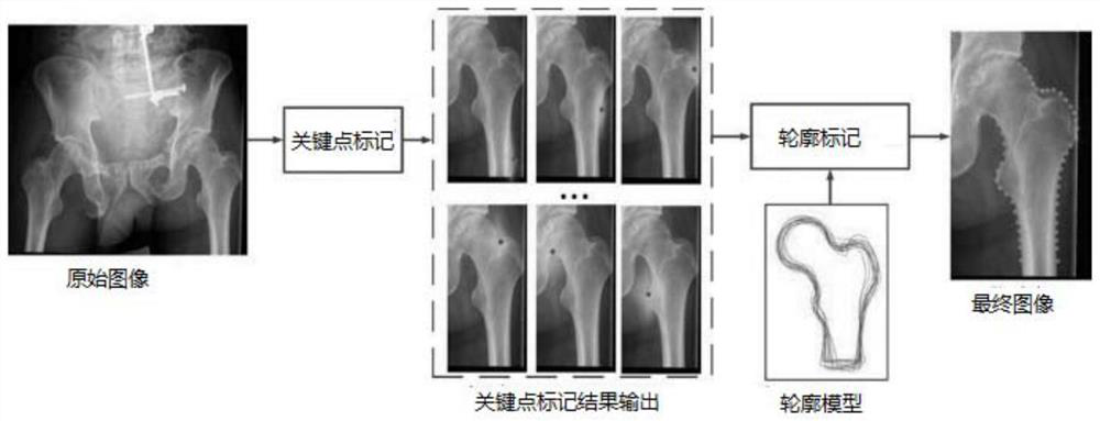
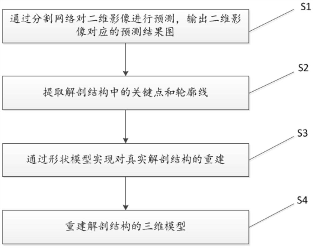
[0070] Such as figure 2 As shown, this embodiment provides a method for automatic recognition of anatomical features and model reconstruction in medical images, including the following steps:

[0071] S1: Input the two-dimensional image to be segmented into the segmentation network for prediction, agree in advance the type of anatomical structure to be recognized, classify and predict each pixel of the input two-dimensional image, and form a prediction result map, wherein, the The segmentation network is obtained by pre-collecting a large number of the two-dimensional images and performing pixel-level labeling, followed by neural network training.

[0072] 1. Predict and mark the 2D image input segmentation network to be segmented

[0073] Specifically, in this embodiment, the purpose of step S1 is to use different labels to predict and mark each different type of anatomical structure in the two-dimensional image, so that the processed prediction results can be clearly ident...

no. 2 example

[0148] The steps of this embodiment are basically the same as those of the first embodiment, the difference is that in step S1, it also includes processing the prediction result graph, specifically:

[0149] 1. Eliminate anxiety

[0150] Perform denoising processing on the prediction result map, and perform connectivity analysis on all pixels classified into this category for all anatomical structure types except the background category. If multiple connected regions are analyzed, find and retain The largest connected region, removing other connected regions.

[0151] Specifically, for the pixels corresponding to each type of label, a largest connected region is found, and other connected regions are deleted. The largest connected region is each type of label, corresponding to an anatomical structure marked.

[0152] 2. Remove jagged points

[0153] Use morphological operations on connected regions to remove small jagged points, as the final prediction result map.

[0154] ...

no. 3 example

[0156] This embodiment combines the methods in the first embodiment and the second embodiment, and uses a specific example to explain the content of the present invention by taking the lower extremity, which accounts for a relatively high proportion in orthopedic clinical research, as an example.

[0157] 1. Training of Segmentation Network

[0158] (1) Collect 90 full-length Dicom images of lower extremities, convert them into 16-bit PNG format after anonymization.


PUM


Abstract

The invention relates to the technical field of medical image intelligent analysis, and provides a medical image anatomical feature automatic identification and model reconstruction method comprisingthe following steps: S1, inputting a two-dimensional image into a segmentation network for prediction, and outputting a prediction result graph corresponding to the two-dimensional image; S2, performing morphological analysis on each anatomical structure segmented from the prediction result graph, and extracting positions and contour lines of key points; S3, reading the contour line and comparingthe contour line with the shape model: when the projection contour simulated by the shape model is close to the contour line, the current shape model is the reconstruction of the real anatomical structure; S4, executing the steps S1-S3 by using at least two two-dimensional images at different visual angles, so as to reconstruct the three-dimensional model of the anatomical structure. According tothe technical scheme, the anatomical feature points and the contour lines of the two-dimensional images can be automatically recognized, and the optimal three-dimensional model can be reconstructed through at least two two-dimensional images at different visual angles.

Description

technical field [0001] The invention relates to the technical field of intelligent analysis of medical images, in particular to a method for automatic recognition of anatomical features in medical images and model reconstruction. Background technique [0002] Two-dimensional images including high-precision X-ray images are very important image references in clinical diagnosis and scientific research. With the help of frontal two-dimensional images, doctors can quantitatively calculate some joint angles and joint distances by manually selecting some anatomical key points and contour lines, and perform alignment analysis, thereby providing preoperative planning for prosthesis replacement surgery and postoperative planning. Provides a numerical basis for evaluating effects. However, manually marking a large number of key points and lines on the image is a great burden for doctors, and the calculation based on the manually marked results is also very time-consuming and labor-in...

Claims


Application Information

Patent Timeline
Patent Type & Authority Applications(China)
IPC IPC(8): G06T7/00G06T17/00G06K9/34G06K9/62G06N3/04
CPCG06T7/0012G06T17/00G06T2207/10081G06T2207/30008G06V10/267G06N3/045G06F18/214
Inventor 朱哲敏蔡宗远
Owner SHANGHAI TAOIMAGE MEDICAL TECH CO LTD
Why Eureka
Try Eureka