Vein closure system

A technology of blood vessel closure and delivery system, applied in the field of medical devices, which can solve problems such as insufficient resolution

Pending Publication Date: 2020-12-11
SHANGHAI SHINEYO MEDICAL (GRP) CO LTD
View PDF0 Cites 3 Cited by
  • Summary
  • Abstract
  • Description
  • Claims
  • Application Information

AI Technical Summary

Problems solved by technology

Despite its popularity, ultrasound visualization provides insufficient resolution when placing catheters intravenousl

Method used


Image

  • Vein closure system
  • Vein closure system
  • Vein closure system

Examples


Embodiment 1

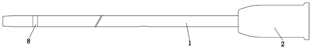
[0051] Embodiment one, such as Figure 1-4 As shown, a vein closure system is composed of a vascular closure glue whose composition is cyanoacrylate and a blood vessel closure glue delivery system, and the blood vessel closure glue delivery system is composed of a catheter 1 and catheter 1 injection parts; inside the catheter 1 There are at least two catheter 1 lumens for providing vascular sealing glue delivery channels, fiber optic channels 22, guide wire channels 24 or ultrasound visible channels 23. The present invention can realize the combined design of different cavities for the catheter 1. Catheter 1 enters the venous blood vessel along the guide wire, and delivers the vascular sealing glue to the blood vessel position through the catheter 1. At the same time, the optical fiber carried inside the catheter 1 can introduce visible light with strong penetrating power into the venous blood vessel. Because the superficial vein is close to the subcutaneous tissue , so the mo...

Embodiment 2

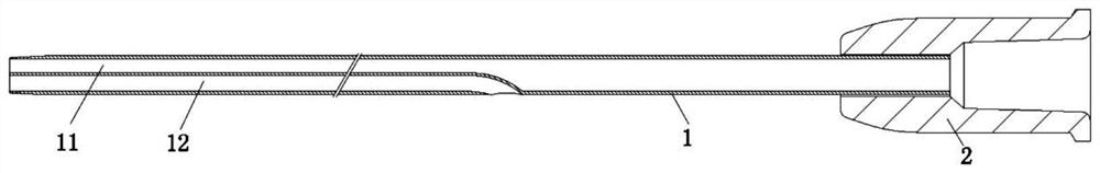
[0060] Embodiment two, such as Figure 5-9 As shown, in this embodiment, the distal end of the second lumen 12 of the catheter is preferably a closed channel, so as to prevent blood from flowing into the second lumen 12 of the catheter along the channel at the distal end of the catheter 1, thereby blocking and polluting the blood in the second lumen 12 of the catheter. The light transmission channel, the distal end of the second cavity 12 of the catheter is preferably sealed with a transparent resin 7 so that the light can pass through the distal end of the catheter 1 smoothly. The proximal end of the second cavity 12 of the catheter is provided with an inlet 3 connected to a light source 4; the light source 4 is preferably visible light with the strongest ability to penetrate skin and blood vessel tissue, preferably red light with the longest wavelength in visible light; the optical fiber is The surface has the quartz optical fiber of polyimide coating, and the optical fiber ...

Embodiment 3

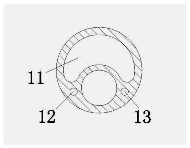
[0062] Embodiment three, such as Figure 10 As shown, in particular, when the second lumen 12 of the catheter is used as a rapid exchange structure, the fiber channel 22 of the second lumen 12 of the catheter and the guide wire channel 24 can be combined into a discontinuous cavity. The increase in the cross-sectional area of ​​the catheter 1 caused by having multiple lumens will lead to poor passability of the catheter 1 in the blood vessel. Therefore, combining the second lumen 12 of the catheter with the fiber optic channel 22 and the guide wire channel 24 into a discontinuous cavity can save the cross-sectional area of ​​the catheter 1; 10 cm is the rapid exchange area of ​​the guide wire of the catheter 1 , and the proximal part is the fiber optic channel 22 .

[0063] Change the catheter 1 into a rapid exchange structure, wherein the blood vessel sealing glue flow channel 21 runs through the entire section of the catheter 1, and the fiber channel 22 and the guide wire c...


PUM


Abstract

The invention belongs to the technical field of medical instruments, and particularly relates to a vein closure system. The vein closure system consists of a blood vessel closing adhesive containing cyanoacrylate and a blood vessel closing adhesive conveying system consisting of a catheter and a catheter injection part; the catheter is internally provided with at least two catheter cavities used for providing a blood vessel closing glue conveying channel, an optical fiber channel, a guide wire channel or an ultrasonic visible channel, different cavities of the catheter are combined for design,the catheter can enter a vein along a guide wire, blood vessel closing glue is conveyed to the position of the blood vessel through the catheter, and besides, an optical fiber carried in the cathetercan introduce visible light with high penetrating power into the vein. The moving positions of the light and the catheter in the superficial vein can be observed due to the fact that the superficialvein is close to subcutaneous tissue, and the catheter can be developed under ultrasound by reasonably selecting the structure and the material of the catheter.

Description

technical field [0001] 本发明属于医疗器械技术领域,特别涉及一种静脉闭合系统。 Background technique [0002] 静脉曲张俗称“蚯蚓腿”或“美腿杀手”,我国的发病率为8.89%至15%之间,粗略统计约有超过一亿静脉曲张患者。下肢静脉曲张是一种血管外科常见疾病,由于在病变初期不痛不痒、没有明显症状,即使样子“丑”一点,很多患者也并不在意。但随着病情发展,“蚯蚓”的存在感会越来越强,引起腿酸、腿沉、腿胀、发痒、疼痛、抽筋、甚至出现皮肤溃疡、破裂、出血等症状。极端情况下,静脉里甚至会形成血栓,严重时引起致命的肺栓塞,也可能造成静脉病变部分破裂造成大出血威胁患者生命安全。 [0003] 静脉曲张是一种临床症状,静脉系统涉及全身各组织器官,上肢静脉回流受阻会出现上肢静脉曲张,肝硬化腹水可能导致腹壁静脉曲张,痔疮其实是痔外静脉丛的淤血曲张,涉及不同的专科,治疗方式也不同。 [0004] 静脉曲张的典型表现是腿上出现像蚯蚓一样的血管,或者蜘蛛纹,静脉曲张的前期症状为小腿酸麻、半夜抽筋、容易感到疲倦、沉重感,随着症状加重,腿上像蚯蚓状的血管则会突出皮肤,甚至形成瘤样的扩张,还能伴随着皮肤瘙痒、湿疹皮炎、色素沉着甚至皮肤溃疡的表现,首选血管外科治疗。 [0005] 目前静脉曲张治疗包括:保守治疗及手术治疗,保守治疗包括:弹力袜治疗及静脉活性药物治疗,手术治疗包括:大隐静脉高位结扎抽剥术、大隐静脉激光治疗术、大隐静脉射频闭合术。还有介于两者之间的硬化疗法。射频消融静脉曲张手术治疗目前最新的微创疗法,是利用射频导管的热效应使曲张静脉迅速纤维化使管腔闭塞,消除静脉曲张。与传统手术相比,创伤更小,几乎无切口,出血更少,更小的副损伤,局部麻醉下完成,术后能立即下地行走活动,日间手术模式。 [0006] 但是,上述手术疗法,比如激光消融、射频消融,包括微波消融等热消融技术,手术前需要在静脉周围注射肿胀剂进行肿胀麻醉,在术后的几天内沿着治疗区域所进行的肿胀麻醉会产生瘀伤和疼痛。射频和激光消融都可能对静脉血管周围的神经产生烧伤或者损伤的副作用,甚至导致手术得不偿失,同时热消融手术除了需要使用昂贵的一次性耗材外,还需要配备昂贵的热发生装置,如射频需要射频发生装置,激光和微波也需要激光源和微波源。 [0007...

Claims


Application Information

Patent Timeline
IPC IPC(8): A61B17/12
CPCA61B17/12109A61B17/12181
Inventor 张娟凤
Owner SHANGHAI SHINEYO MEDICAL (GRP) CO LTD
Why Eureka
Try Eureka