Bone surface segmentation method under ultrasonic imaging

An ultrasound imaging and surface segmentation technology, which is applied in image analysis, image enhancement, image data processing, etc., can solve the problem that the algorithm performance is difficult to meet the real-time requirements of the imaging system, and achieve high-speed segmentation, improved real-time performance, and high precision. Effect

Pending Publication Date: 2020-12-18
HARBIN ENG UNIV
View PDF11 Cites 1 Cited by
  • Summary
  • Abstract
  • Description
  • Claims
  • Application Information

AI Technical Summary

Problems solved by technology

However, the existing convolutional neural network-based methods basically need to preprocess the ultrasound images to varying degrees, and this process takes an average of 1 to 2 seconds per image, and then segment after preprocessing, which leads to The performance of the algorithm is difficult to meet the real-time requirements of the imaging system in the CAOS system

Method used


Image

  • Bone surface segmentation method under ultrasonic imaging
  • Bone surface segmentation method under ultrasonic imaging
  • Bone surface segmentation method under ultrasonic imaging

Examples


Embodiment approach

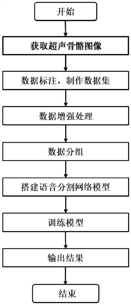
[0040]This method is based on a convolutional neural network to achieve segmentation and extraction of bone indications under ultrasound images. The following will further describe the implementation method of the present invention in detail in conjunction with specific embodiments as follows:

[0041]1. Hardware configuration environment

[0042]The hardware used in the present invention includes: (1) a computer with an image processing card (2080Ti), (2) Mindray, DP10 portable ultrasonic imager.

[0043]2. Software configuration environment

[0044]Python, Pytorch, openCV

[0045]3. Such asfigure 1 As shown, the present invention proposes a deep learning-based ultrasound image segmentation method for bone indication, which specifically includes the following steps:

[0046]Step 1: Use an ultrasound probe to obtain ultrasound pictures of the bone surface, and clean the obtained images (for example, delete pictures with poor imaging quality).

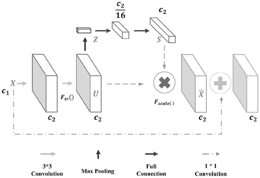
[0047]Step 2: Perform center cropping on the original ultra...


PUM


Abstract

The invention provides a bone surface segmentation method under ultrasonic imaging. The method is characterized in that the method is realized by using a python language, and comprises the following steps: using an ultrasonic probe for acquiring an ultrasonic picture of a bone surface, and cleaning the acquired image; carrying out center cutting on the original ultrasonic image to obtain an ultrasonic imaging area with the size of 288*320, and removing useless areas; marking the bone ultrasonic image to make a mask mark image, thereby making a data set; enhancing the data set obtained in the step 3 by using an image enhancement technology, enriching data contents, and expanding the number of samples; randomly and equally dividing the data set into five parts; building a neural network model, which is composed of two modules, namely an encoding module and a decoding module; and performing five-fold cross validation training on the network model by utilizing the five data sets obtained in the step 5. The system can be integrated into a universal computer-aided system with the help of a common camera, extra equipment is not needed, and the system is suitable for hospitals and families.

Description

Technical field[0001]The invention relates to a bone surface segmentation method, in particular to a bone surface segmentation method under ultrasound imaging, and belongs to the computer vision field of a computer-assisted surgical navigation system.Background technique[0002]Fracture is a common surgical disease in clinic, which seriously affects people's health and life. With the increase in the types of outdoor sports, there are more and more sports traumas, and the increase in the incidence of traffic accidents also gradually increases the number of fracture patients.[0003]Orthopedic surgery is an important solution for the treatment of fractures. The use of high-precision intraoperative navigation technology to guide the operation during the operation is a key to reducing the sequelae of postoperative recovery of fracture patients. For the imaging and positioning of the fractured area, the computer-assisted orthopedic surgery system (CAOS) is an effective auxiliary method, and ...

Claims


Application Information

Patent Timeline
Patent Type & Authority Applications(China)
IPC IPC(8): G06T7/11G06T3/60G06T5/00
CPCG06T7/11G06T3/60G06T5/003G06T2207/20081G06T2207/20084G06T2207/10132G06T2207/20132G06T2207/30008
Inventor 栾宽李泽钰李金刘小龙周洋王鹏
Owner HARBIN ENG UNIV
Why Eureka
Try Eureka