
Scanning electron microscope sample table and sample preparation method thereof

A scanning electron microscope and sample stage technology, applied in the direction of circuits, discharge tubes, electrical components, etc., can solve problems such as not easy to fix, break, and easily deform, and achieve the effect of protecting the electron microscope, improving contact, and ensuring that it is not damaged

Pending Publication Date: 2021-01-12
DALIAN UNIV OF TECH
View PDF0 Cites 0 Cited by
  • Summary
  • Abstract
  • Description
  • Claims
  • Application Information

AI Technical Summary

Problems solved by technology

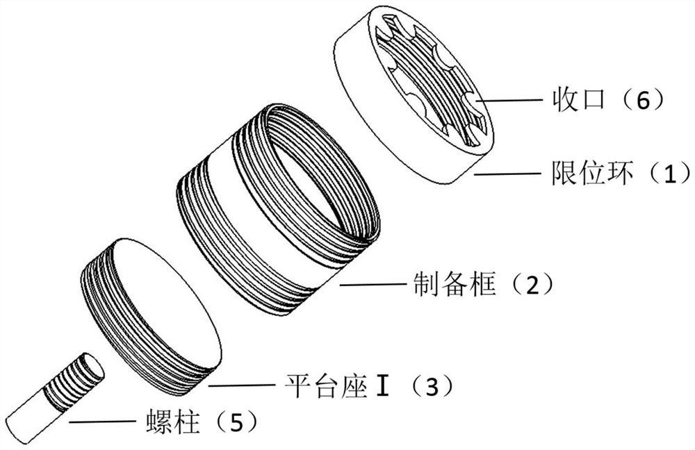
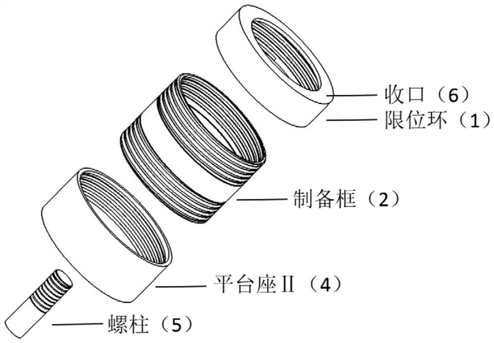
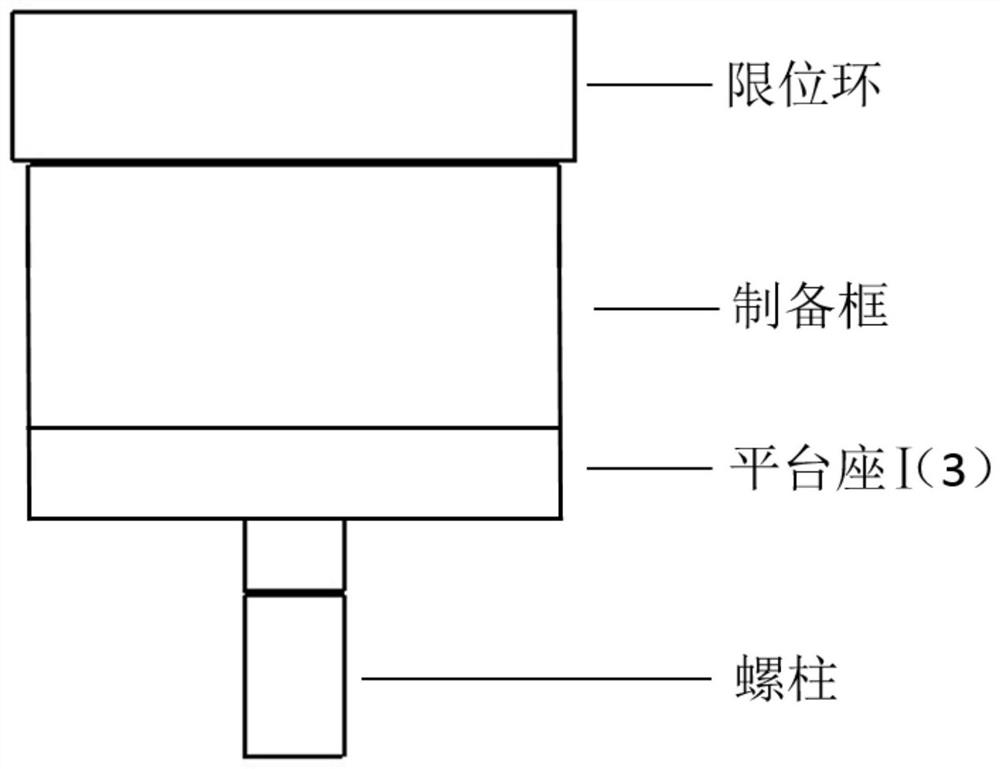
[0004] Aiming at the technical problem that the fragile freeze-dried macroporous material is not easy to be fixed on the scanning electron microscope sample stage, and is easily deformed and broken, the present invention provides a scanning electron microscope sample stage and its sample preparation for the macroporous monolithic material obtained by the freeze-drying method Method, by splitting the sample stage into three main parts: the limit ring, the preparation frame and the platform seat, according to the volume shrinkage of the sample after freeze-drying and the performance of the surface pore structure, these three parts are assembled and combined flexibly and rationally , to achieve good contact and fixation of the sample in the sample stage, and to optimize the pore structure of the sample surface

Method used


Image

  • Scanning electron microscope sample table and sample preparation method thereof
  • Scanning electron microscope sample table and sample preparation method thereof
  • Scanning electron microscope sample table and sample preparation method thereof

Examples


Embodiment 1

[0040] The limiting ring 1, the preparation frame 2, the platform seat I3 and the stud 5 are made of metallic copper.

[0041] The outer diameter of the limit ring 1 is 36mm, the inner diameter is 34mm, the mouth 6 is 8 claws, the claws protrude 3mm toward the center of the circle, the width of the claws is 2mm, and the thickness of the mouth is 1mm.

[0042] The inner diameter of the preparation frame 2 is 30mm, the outer diameter is 34mm, and the height is 20mm. The width of the thread at the upper and lower ends of the outer side is 2mm, and the width of the threaded opening at the upper and lower ends of the inner side is 6mm. The outer thread and the limit ring are assembled.

[0043] The thickness of the platform base I3 is 4mm, the diameter is 30mm, the inner diameter of the small hole in the bottom center is 3.2mm, and the depth of the internal thread is 3mm. The external thread on the edge is screwed into the assembly from one end of the preparation frame.

[0044] Th...

Embodiment 2

[0048] The limiting ring 1, the preparation frame 2, the platform seat I3 and the stud 5 are made of aluminum.

[0049] The outer diameter of the limit ring 1 is 16 mm, the inner diameter is 14 mm, the limit ring closing 6 is circular, the ring width is 2 mm, and the thickness of the closing part is 0.5 mm.

[0050] The inner diameter of the preparation frame 2 is 10mm, the outer diameter is 14mm, and the height is 10mm. The width of the external thread at the upper and lower ends of the outer side is 2mm, and the width of the inner thread at the upper and lower ends of the inner side is 3mm. The outer thread at one end is assembled with the limit ring 1.

[0051] The platform seat I3 has a thickness of 3 mm, a diameter of 10 mm, an inner diameter of the small hole in the bottom center of 3.2 mm, and an internal thread depth of 2 mm. The external thread on the outer edge of the platform is screwed in from one end of the preparation frame 2 for assembly.

[0052] The diameter o...

Embodiment 3

[0056] The limit ring 1, the preparation frame 2, the platform seat I3, the platform seat II4 and the stud 5 are made of metallic copper.

[0057] The outer diameter of the limiting ring 1 is 26 mm, the inner diameter is 24 mm, the closing 6 of the limiting ring 1 is 4 claws, the claws protrude 1 mm toward the center of the circle, the width of the claws is 1 mm, and the thickness of the closing part is 1 mm.

[0058] The inner diameter of the preparation frame 2 is 20mm, the outer diameter is 24mm, and the height is 15mm. The width of the external thread at the upper and lower ends of the outer side is 2mm, and the width of the inner thread at the upper and lower ends of the inner side is 5mm. The outer thread at one end is assembled with the limit ring 1.

[0059] The platform seat I3 has a thickness of 4mm, a diameter of 20mm, an inner diameter of the bottom center hole of 3.2mm, and an internal thread depth of 3mm. The external thread on the outer edge is screwed in from on...


PUM

PropertyMeasurementUnit
thicknessaaaaaaaaaa
heightaaaaaaaaaa
heightaaaaaaaaaa

Abstract

The invention provides a scanning electron microscope sample table for a macroporous integral material obtained by a freeze drying method and a sample preparation method thereof. Sample preparation and electron microscope sample preparation are completed in one step through in-situ forming of a macroporous material in the sample table, crushing and deformation of a fragile macroporous integral material caused by cutting, transferring and fixing during sample preparation of a scanning electron microscope are avoided, good fixation and protection are provided for a sample, a macroporous structure of the sample is prevented from being damaged, and an effect of protecting an electron microscope is achieved. The sample table is divided into three main components, namely a limiting ring, a preparation frame and a platform base, and the three components can be flexibly and reasonably assembled and combined according to the volume shrinkage of the sample and the presentation condition of a surface pore structure, so that the contact between the sample and the sample table is enhanced, the optimization of the surface pore structure of the sample is realized, the electron microscope sample preparation is more efficient, and test results are more ideal.

Description

technical field [0001] The invention relates to the technical field of scanning electron microscope sample preparation, in particular to a scanning electron microscope sample stage and a sample preparation method for macroporous monolithic materials obtained by a freeze-drying method. Background technique [0002] It is a common and irreplaceable method to observe the morphology of macroporous monolithic materials obtained by freeze-drying method by scanning electron microscope, but many macroporous materials prepared by freeze-drying method have poor strength, easy to break, and easy to deform. There are often great difficulties in the preparation of SEM samples. [0003] Although the sample preparation of the scanning electron microscope is simple, the sample must be firmly fixed on the sample stage, otherwise the sample is prone to charging phenomenon due to poor conductivity, and the fallen sample or debris will pollute the sample chamber of the electron microscope, brin...

Claims


Application Information

Patent Timeline
Patent Type & Authority Applications(China)
IPC IPC(8): G01N23/2202H01J37/20
CPCG01N23/2202H01J37/20
Inventor 万鹏
Owner DALIAN UNIV OF TECH
Learn More