Application of Ptbp1 inhibitor in preventing and/or treating nervous system diseases related to functional neuron death
A neurological disease, neuron technology, applied in the field of biomedicine, can solve problems such as failure to prevent the development of the disease
- Summary
- Abstract
- Description
- Claims
- Application Information
AI Technical Summary
Problems solved by technology
Method used
Image
Examples
Embodiment 1
[0304] Example 1 Using CasRx to specifically knock down Ptbp1 in vitro
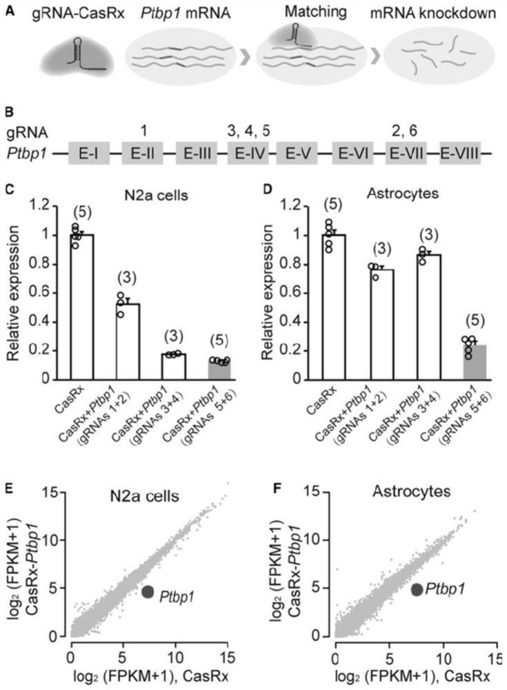
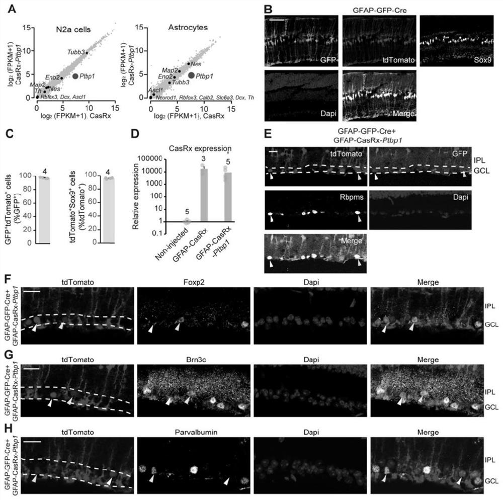
[0305] To determine the efficiency of CasRx-mediated Ptbp1 knockdown, six gRNAs targeting Ptbp1 were first designed, and their inhibitory efficiencies were compared in cultured N2a cells and astrocytes ( figure 1 A and 1B). It was found that co-transfection of two gRNAs (combination of 5 and 6) targeting Ptbp1 exon IV and VII regions could achieve 87% ± 0.4% and 76% ± 4% in N2a cells and astrocytes, respectively. decreased (Figure 1C and 1D). RNA whole-transcriptome sequencing data further showed that this knockdown was very specific ( figure 1 E, 1F and 2A).
Embodiment 2
[0306] Example 2 Conversion of Müller glia to optic ganglion cells in the mature retina
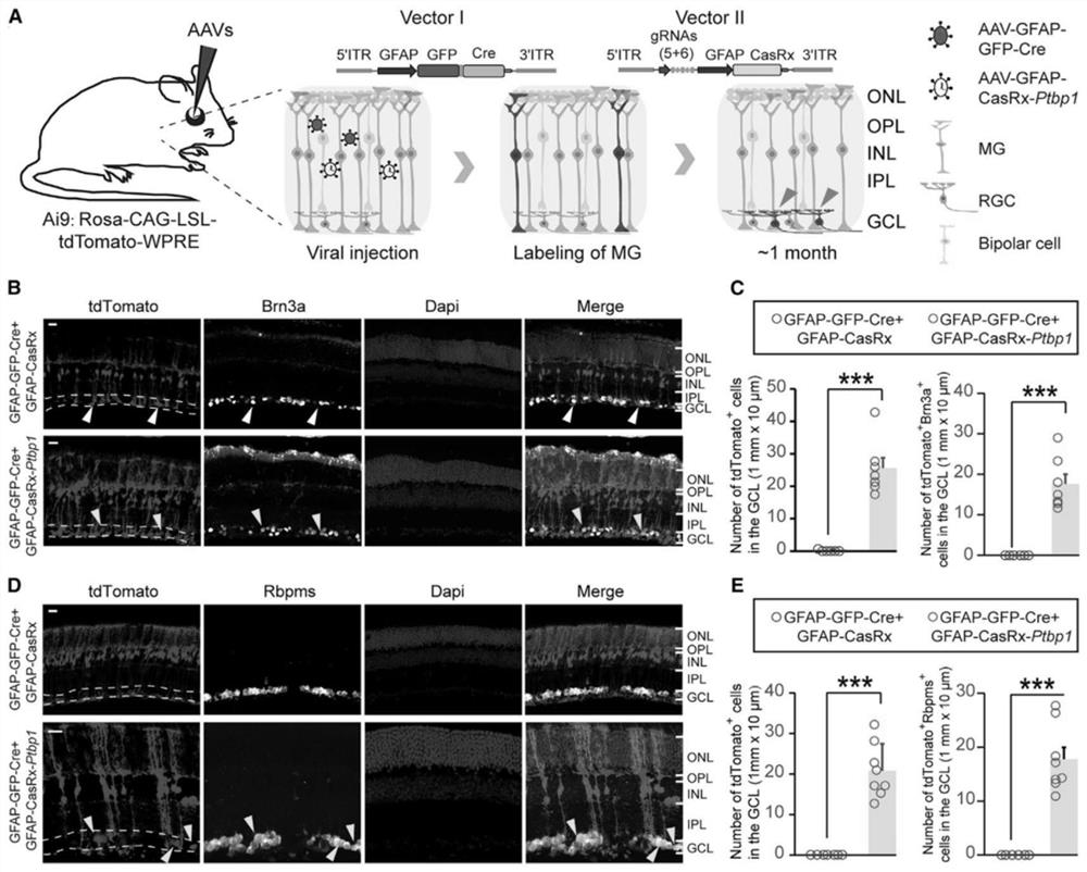
[0307] Previous studies have found that knockdown of Ptbp1 by shRNA can convert cultured mouse fibroblasts and N2a cells into functional neurons, and next investigated whether Ptbp1 knockdown in the mature retina regenerates Müller glial cells in vivo. into optic ganglion cells. To specifically and permanently label retinal Müller glia, we injected AAV-GFAP-GFP-Cre into the eyes of Ai9 mice (Rosa-CAG-LSL-tdTomato-WPRE) to specifically induce tdTomato in Müller glia Expression in cells ( figure 2 B and 2C). We also constructed AAV-GFAP-CasRx-Ptbp1 (gRNA 5+6) driven by the Müller glial cell-specific promoter GFAP, hoping to specifically knock down Ptbp1 in Müller glial cells and we simultaneously constructed A control virus AAV-GFAP-CasRx not targeting Ptbp1 ( image 3 A and 2D). We first co-injected AAV-GFAP-CasRx-Ptbp1 and AAV-GFAP-Cre-GFP under the retina of Ai9 mice aged about 5 w...
Embodiment 3
[0308] Example 3 Conversion of MG to RGC in retinal injury mouse model
[0309] To explore whether MG-induced RGCs could replenish lost RGCs in a mouse model of retinal injury, we injected NMDA (200 mM) intravitreally into 4- to 8-week-old Ai9 mice, resulting in most Death of RGCs and reduction of inner plexiform layer (IPL) thickness. Two to three weeks after injection of NMDA, AAV-GFAP-CasRx-Ptbp1 plus AAV-GFAP-GFP-Cre or control AAV virus ( Figure 6 A and 6B). One month after AAV injection, we found that the number of Brn3a or Rbpms-positive cells in the optic ganglion cell layer was significantly increased in AAV-GFAP-CasRx-Ptbp1-injected retinas. Interestingly, most of these cells were positive for tdTomato. However, we did not find an increase in the optic ganglion cell layer injected with control AAV ( Figure 6 C-6F). To determine whether MG-induced RGCs are integrated into retinal circuits and have the ability to receive visual information, we performed extracel...
PUM
| Property | Measurement | Unit |
|---|---|---|
| thickness | aaaaa | aaaaa |
Abstract
Description
Claims
Application Information
 - R&D
- Intellectual Property
- Life Sciences
- Materials
- Tech Scout
- Unparalleled Data Quality
- Higher Quality Content
- 60% Fewer Hallucinations
Browse by: Latest US Patents, China's latest patents, Technical Efficacy Thesaurus, Application Domain, Technology Topic, Popular Technical Reports.
© 2025 PatSnap. All rights reserved.Legal|Privacy policy|Modern Slavery Act Transparency Statement|Sitemap|About US| Contact US: help@patsnap.com


