Renal tubule detection and segmentation method based on UNET

A renal tubule and tissue technology, applied in the field of image detection, recognition and segmentation, can solve problems such as no technical significance, inability to segment renal tubules, and no mention of renal tubule-related content, and achieve the effect of improving automation and accuracy.

Active Publication Date: 2021-03-16
杭州医派智能科技有限公司
View PDF9 Cites 5 Cited by
  • Summary
  • Abstract
  • Description
  • Claims
  • Application Information

AI Technical Summary

Problems solved by technology

Therefore, in the application scenario of digital film reading, the renal tubules in the same image cannot be well segmented only by traditional image recognition and model algorithms
[0006] In the prior art, there are few targeted literature reports in this regard, and the research in the prior art mainly focuses on the imaging research. For example, the Chinese patent application CN 108885204A discloses a method for predicting heterogeneous A high-throughput imaging-based approach to cell-type-specific toxicity of organisms that provides a means to predict in vivo proximal tubular, bronchial epithelial, and alveolar cell-specific toxicity of soluble or particulate compounds with virtually no And about the related content of renal tubule in image segmentation, there is no technical significance for the problems mentioned in the background technology of the present invention

Method used


Image

  • Renal tubule detection and segmentation method based on UNET
  • Renal tubule detection and segmentation method based on UNET
  • Renal tubule detection and segmentation method based on UNET

Examples


Embodiment 1

[0068] Embodiment 1, with reference to attached Figure 1-9 .

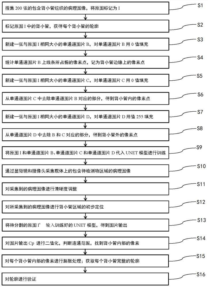
[0069] The present invention provides a method for detecting and segmenting renal tubules based on UNET. The detecting and segmenting method is used to detect and segment renal tubule regions in collected pathological images. The detecting and segmenting method includes a training stage and an inference stage. The training stage is used to mark the renal tubule data and train the UNET model, and the reasoning stage uses the trained UNET model for the detection and segmentation of renal tubular regions.

[0070] Specifically, as figure 1 As shown, the detection segmentation method of the present invention comprises the following steps:

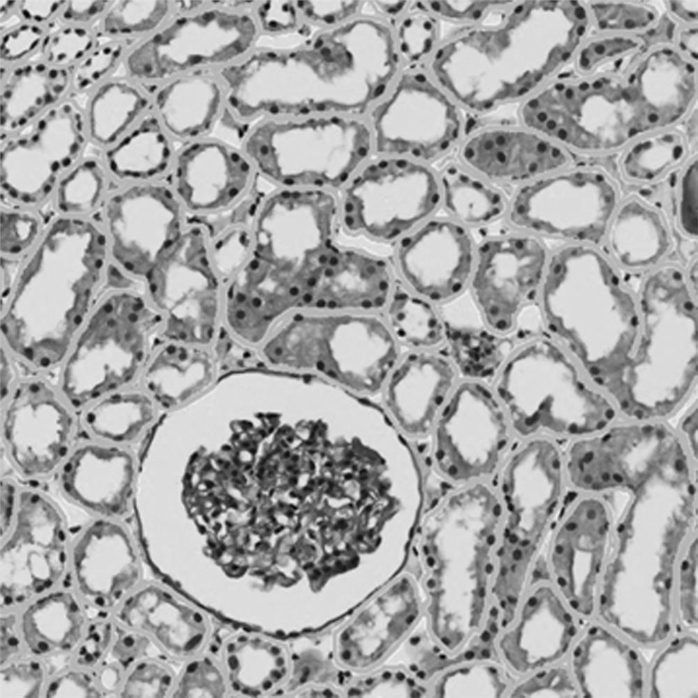
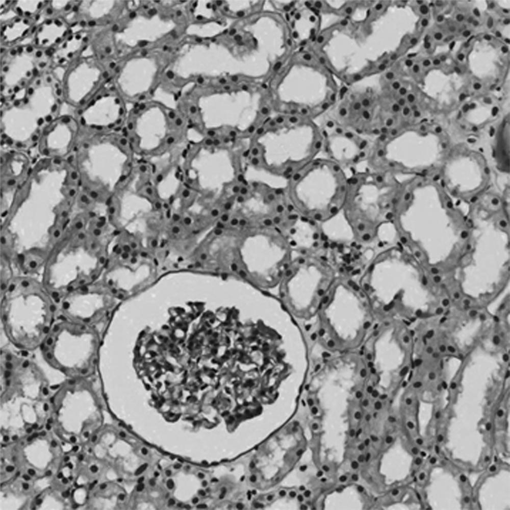
[0071] S1, collect 200 pathological images containing renal tubular tissue, and mark the original images as I, such as figure 2 As shown, for the convenience of description, one of the original drawings I is taken as an example below for illustration.

[0072] S2, use the open...

Embodiment 2

[0104] Embodiment 2, with reference to attached Figure 10 .

[0105] In this embodiment, a computer device 100 is provided, including a memory 101, a processor 102, and a computer program 103 stored in the memory 101 and operable on the processor 102. When the processor 102 executes the computer program 103, it can realize The steps in the detection and segmentation method provided by the above-mentioned embodiment 1.

Embodiment 3

[0107] In this embodiment, a computer-readable storage medium is provided, on which a computer program is stored, and when the computer program is executed by a processor, the steps in the detection and segmentation methods provided in the above-mentioned embodiments can be implemented.

[0108] In this embodiment, the computer program may be the computer program in Embodiment 2.

[0109] In this embodiment, the computer-readable storage medium may be executed by the computer device in Embodiment 2.


PUM


Abstract

The invention provides a renal tubule detection and segmentation method based on UNET, which is used for detecting and segmenting an area where renal tubule tissue is located in a collected pathological image, and comprises a training stage and an inference stage; the training stage is used for marking renal tubule data, and training a UNET model; and the inference stage is used for applying the trained UNET model to detection and segmentation of renal tubular tissues in a pathological image. According to the segmentation method, the area corresponding to the renal tubular tissue in the pathological strip image can be accurately recognized, the adjacent renal tubular tissue can be well separated, digital film reading is facilitated, and the automation and accuracy of digital film reading are improved.

Description

technical field [0001] The present invention relates to the technical field of image detection, recognition and segmentation, more specifically, it relates to a method for identifying and separating a characteristic pathological region in a pathological strip. Background technique [0002] Pathological examination is a pathomorphological method used to examine pathological changes in body organs, tissues or cells. In order to explore the disease process of organs, tissues or cells, some pathological and morphological examination method can be used to check the lesions they have, discuss the causes of lesions, pathogenesis, and the development process of lesions, and finally make a pathological diagnosis. diagnosis. The examination method of pathomorphology first observes the pathological changes of the gross specimen, then cuts out a certain size of lesion tissue, makes pathological sections with histopathological method, and further examines the lesion with a microscope. ...

Claims


Application Information

Patent Timeline
Patent Type & Authority Applications(China)
IPC IPC(8): G06T7/00G06T7/11G06T7/13G06T7/181G06T7/187G01N21/84
CPCG06T7/0012G06T7/11G06T7/13G06T7/181G06T7/187G01N21/84G06T2207/10056G06T2207/20081G06T2207/30024G06T2207/30084
Inventor 汪太平张敏飞
Owner 杭州医派智能科技有限公司
Why Eureka
Try Eureka