
Joint segmentation method for optic cups and optic disks in fundus medical images

A joint segmentation and medical imaging technology, applied in the field of medical image processing, can solve the problems of insufficient segmentation network context information extraction, blurred boundaries, insufficient multi-scale information extraction, etc., and achieve the effect of improving the efficiency and quality of joint segmentation.

Pending Publication Date: 2021-04-02
SUZHOU UNIV
View PDF6 Cites 4 Cited by
  • Summary
  • Abstract
  • Description
  • Claims
  • Application Information

AI Technical Summary

Problems solved by technology

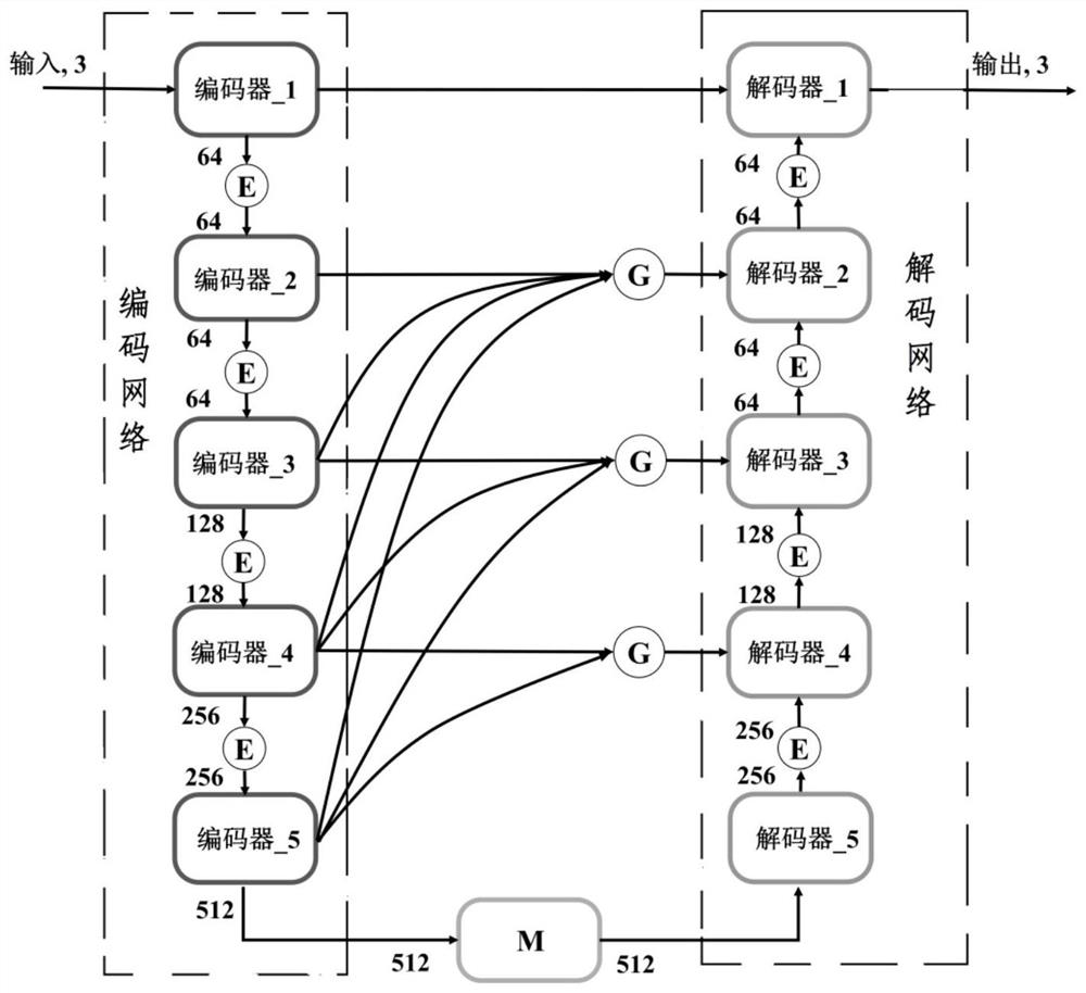
U-Net adopts the structure of encoder and decoder, combined with skip connection, which has greatly improved the accuracy of medical image segmentation, but the segmentation network is still not enough to extract the context information of the image, which will cause the network to be difficult for the image. The multi-scale information extraction is not enough, and it is easy to lose the detailed feature information; in addition, its skip connection only transfers the feature information from the encoder to the decoder, ignoring the use of global information, which leads to the network pair with blurred boundaries and low contrast. When the target is segmented, it is prone to mis-segmentation
[0007] Therefore, the existing medical image segmentation methods have shortcomings such as insufficient extraction of the global context information and multi-scale context information of the fundus color photo image, which affects the segmentation effect and cannot meet the segmentation requirements of the cup and disc in fundus medical images.

Method used


Image

  • Joint segmentation method for optic cups and optic disks in fundus medical images
  • Joint segmentation method for optic cups and optic disks in fundus medical images
  • Joint segmentation method for optic cups and optic disks in fundus medical images

Examples


Embodiment Construction

[0029] The present invention will be further described below in conjunction with the accompanying drawings and specific embodiments, so that those skilled in the art can better understand the present invention and implement it, but the examples given are not intended to limit the present invention.

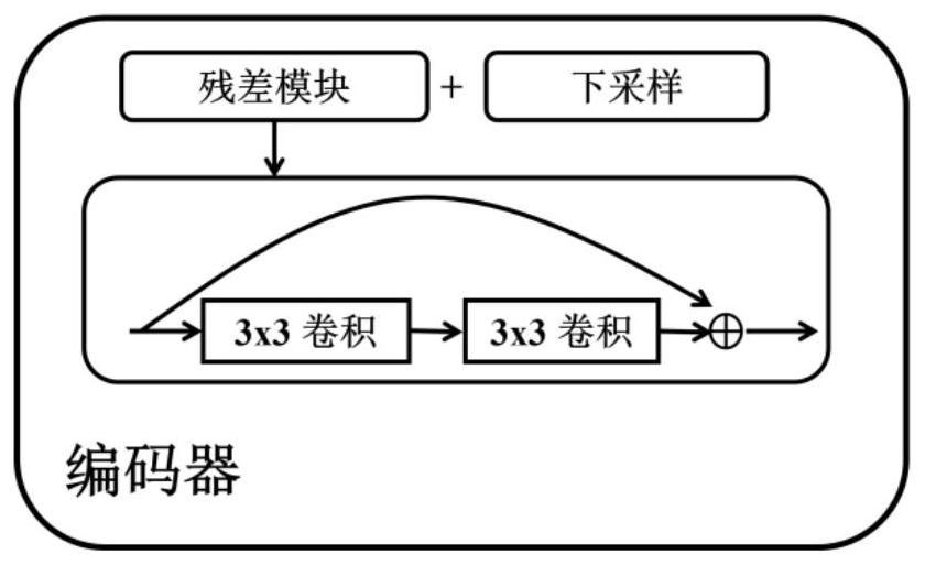
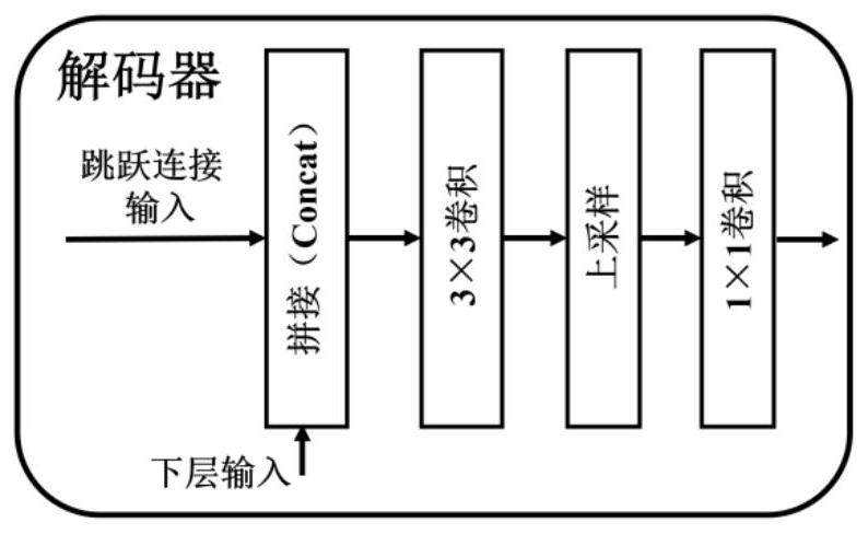
[0030] refer to Figure 1-Figure 3 , figure 1 middle Indicates the global information extraction module - GCE module, Denotes the channel attention module, Representing the multi-path atrous convolution module-MAC module, this embodiment discloses a method for joint segmentation of optic cup and optic disc in fundus medical images, including the following steps:

[0031] S1) Construct a joint segmentation network based on the U-Net network;

[0032] The U-Net network includes an encoding network and a decoding network. The encoding network includes multiple encoders. The decoding network includes multiple decoders. The encoders and decoders correspond one-to-one. Each encod...


PUM


Abstract

The invention discloses a joint segmentation method for optic cups and optic disks in fundus medical images, which comprises the following steps: firstly, constructing a joint segmentation network based on a UNet network, and introducing a global information extraction module and a multi-path hole convolution module into the joint segmentation network; and inputting a fundus medical image to be processed into the joint segmentation network for joint segmentation of the optic cups and the optic disks. According to the method, global context information and multi-scale context information in thefundus medical image can be fully extracted, and the joint segmentation effect of optic cups and optic disks in the fundus medical image is improved.

Description

technical field [0001] The invention relates to the technical field of medical image processing, in particular to a joint segmentation method of optic cups and optic discs in fundus medical images. Background technique [0002] Medical imaging refers to the technology and processing process of obtaining internal tissue and organ images of the human body or a certain part of the human body in a non-invasive manner for the purpose of medical treatment or medical research. According to different implementation steps, medical imaging includes medical imaging technology and medical processing technology. With the help of medical imaging technology, medical personnel can have a clearer understanding of the specific tissue and organ conditions of the human body and then provide more accurate and reasonable diagnosis and treatment plans. Medical image segmentation is a key technology in modern medical image processing, and it is the basis for follow-up operations such as 3D reconstr...

Claims


Application Information

Patent Timeline
IPC IPC(8): G06T7/00G06T7/10
CPCG06T7/0012G06T7/10G06T2207/30041G06T2207/20081G06T2207/20084G06T2207/20132
Inventor 朱伟芳朱乾龙陈新建
Owner SUZHOU UNIV
Learn More